Wikipedia:Featured picture candidates/Digestive system diagram
Digestive system diagram edit
Voting period is over. Please don't add any new votes. Voting period ends on 22 Oct 2015 at 19:07:14 (UTC)

- Reason
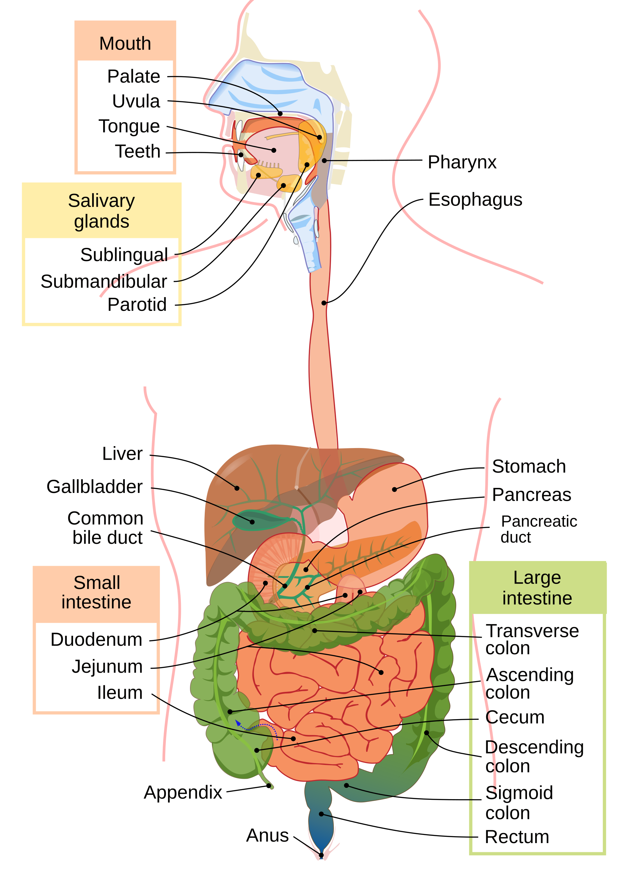
- High EV. High quality. Featured Picture on Commons. Picture of the Day on Commons.
- Articles in which this image appears
- Gastrointestinal physiology, Ascending colon, Transverse colon, Descending colon, Salivary gland, Submandibular gland, Sublingual gland, Ethanol
- FP category for this image
- Wikipedia:Featured pictures/Sciences/Biology
- Creator
- LadyofHats
- Support as nominator – — Cirt (talk) 19:07, 12 October 2015 (UTC)
- Support adds positively (as an image/diagram)--Ozzie10aaaa (talk) 19:53, 12 October 2015 (UTC)
- Thank you for your comments and your Support, Ozzie10aaaa, much appreciated. — Cirt (talk) 20:11, 12 October 2015 (UTC)
- Note: Threaded discussion and back-and-forth moved to talk page.
- I changed the rule to make this case easier to evaluate. Support based on meeting the criteria for resolution and for being a useful image. Blue Rasberry (talk) 20:46, 12 October 2015 (UTC)
- Note: This file has annotations. Move the mouse pointer over the image to see them. Please also see higher resolution here and 2000px here. Thank you, — Cirt (talk) 20:19, 12 October 2015 (UTC)
- Again, just to reiterate, per the featured picture criteria, vector graphics in SVG can be infinitely scaled without loss of quality. Note the word INFINITELY. Surely INFINITE size with zero loss of quality is a high enough picture size. Thank you. — Cirt (talk) 20:52, 12 October 2015 (UTC)
- Support SVG can be scaled infinitely, so per the FPC this meets or exceeds minimum resolution requirements. RO(talk) 21:06, 12 October 2015 (UTC)
- Support - SchroCat (talk) 21:13, 12 October 2015 (UTC)
- Comment Beautiful picture, but some of the spatial relationships are incorrect. In lateral projection with the head rotated like this, the parotid gland would overlap rather than lie posterior to (i.e. to the right in this cartoon) the posterior orpharynx. Also, the esophagus enters the abdomen and connects to the cardia and fundus of the stomach posterior to the left lobe of the liver. soupvector (talk) 21:44, 12 October 2015 (UTC)
- Thank you, Soupvector, for the comment. I still feel it's quite high quality and the entrance is likely being shown as such for illustrative purposes. — Cirt (talk) 21:47, 12 October 2015 (UTC)
- Further, for the basic purpose of illustrating names of key structures in the digestive system, this image is both of High Quality (as demonstrated by Picture of the Day on Commons), and High Encyclopedic Value (as demonstrated by its use already on wiki articles as noted, above.) Thank you, — Cirt (talk) 22:39, 12 October 2015 (UTC)
- Oppose until inaccuracies are addressed. One of our criteria is accuracy and verifiability. Misrepresenting the subject is an issue. Also, aren't there higher quality illustrations from books like Sobotta's Atlas and Text-book of Human Anatomy? Something like this would work much better than the fairly simple SVG we see here. — Chris Woodrich (talk) 23:30, 12 October 2015 (UTC)
- Once again, it is Featured Picture on Commons. Picture of the Day on Commons. Already used in articles Gastrointestinal physiology, Ascending colon, Transverse colon, Descending colon, Salivary gland, Submandibular gland, Sublingual gland. High Quality as determined by Commons. High EV as already used in High Encyclopedic value in multiple articles on medicine. Thank you, — Cirt (talk) 23:37, 12 October 2015 (UTC)
- Note: Please note author's explanations at successful Commons promotion to Featured Picture discussion. The graphic was created to emphasize the digestive system and the key labeled organs -- note that there are annotations. Move the mouse pointer over the image to see them. — Cirt (talk) 23:44, 12 October 2015 (UTC)
- Oppose this is a very high-quality image used on many articles that adds significantly to understanding, and I'm very grateful to the creator for making it. However I'm very uncomfortable promoting it to a "featured picture" because it's factually incorrect, something I feel should be a determining characteristic of featured pictures. Some examples:
- Nasal cavity bottom should be flat
- Uvula or soft palate?
- Epiglottis not labelled
- Abdominal contents quite high, as discussed
- Esophagus should be behind liver, as discussed
- Shape of stomach does not show pylorus
- Small intestine appears to be behind ascending colon
- Atypical labelling of common bile duct, this usually points to the duct above and the ampulla of Vater is usually where the label currently lies
- Ascending colon arrow points to the Taenia coli but descending to a haustra.
- We may not have many anatomical featured images and so be it, I think the factual standards for accuracy should apply here just as they do in GA and FA. So without trying to diminish the high quality of this work, I appose this nomination for its accuracy. This oppose stands regardless of what commons users thought in 2007. --Tom (LT) (talk) 10:57, 13 October 2015 (UTC)
- (E/C) Oppose due to inaccuracies and thus diminished EV, per soupvector and Chris Woodrich. Here are some further concerns:
- The style is inconsistent. The intestines are shown as exteriors, while the stomach is shown as a hollow cross-section. Furthermore, the stomach's walls are shown in the same colour and confusingly appear continuous with the entire oesophagus and the nasal cavity and phraynx, which are empty spaces. Also, the highlighted colour of the oral cavity loses the fact that it is continuous with the pharynx.
- It isn't clear what the shading in the liver is supposed to show. At any rate, it doesn't help convey that the gallbladder and bile ducts attach to the "underside" of the liver which is to the back. As with the liver/stomach positions, the entire image needs a better way of representing front and behind, not just simply changing the anatomy, which is misleading.
- If the anatomical distortions are deliberate, in order to show each structure unobscured, this needs to be properly conveyed.
- The roof of the mouth is far too thick.
- --Paul_012 (talk) 11:08, 13 October 2015 (UTC)
- in regard to your second point it does seem as if the intention is to show the anatomical underlyings...--Ozzie10aaaa (talk) 15:02, 13 October 2015 (UTC)
- Oppose Sorry for that, because it looks so well, but the criticism concerning the content seems to have a broad base. --Tremonist (talk) 14:51, 13 October 2015 (UTC)
- WITHDRAW PLEASE. This does not have a chance of passing and it's not worth proceeding. Thank you for your comments, above. — Cirt (talk) 15:08, 13 October 2015 (UTC)
Not Promoted --Armbrust The Homunculus 15:22, 13 October 2015 (UTC)
- Withdrawn nomination. Armbrust The Homunculus 15:22, 13 October 2015 (UTC)