| Mr. Ibrahem/Chest tube | |
|---|---|
 The free end of the chest tube is usually attached to an underwater seal, below the level of the chest. This allows the air or fluid to escape, but prevents anything returning to the chest. | |
| Other names | Intercostal drain, chest drain, thoracic catheter, tube thoracostomy, intercostal catheter, Bülau drain |
| Specialty | Emergency medicine, general surgery |
| Complications | Subcutaneous emphysema, blockage of the tube, bleeding, infection, reexpansion pulmonary edema, lung or diaphragm injury[1] |
A chest tube is a flexible plastic tube that is inserted through the chest wall into the pleural space.[1] It is used to remove air (pneumothorax), fluid (pleural effusion, hemothorax), or pus (empyema).[1] In those with a tension pneumothorax, a needle thoracostomy may be carried out first.[1]
The person is generally placed on their back, possibly with the head of the bed raised, and their hand behind their head.[2] After the area is cleaned with chlorhexidine and injected with local anesthetic, a 2 cm horizontal incision is make at the 4th or 5th intercostal space anterior axillary line.[2] Kelly forceps are than used to dissect one intercostal space up and into the pleura.[2] A finger may be placed in the hole to help guide in the chest tube.[2] The tube is aimed superiorly for air and inferiorly for fluid.[2] The tube is than sutured, gauze applied, and taped.[2] It is than connected to a drainage system.[2]
In a pneumothorax bubbles will be seen in the drainage system.[2] A chest X-ray is than carried out to confirm placement.[2] The tube may be removed once air leakage has stopped for more than 12 to 24 hours or the amount of fluid out is under 200 ml per 24 hours.[2] Removal should occur when the person has taken and is holding a maximal breath.[2] Another chest X-rays is done after 12 to 24 hours.[2] Complications may include subcutaneous emphysema, blockage of the tube, bleeding, infection, reexpansion pulmonary edema, and lung or diaphragm injury.[1]
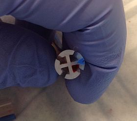
A chest tube was first used by Hippocrates around 400 BC to drain an empyema using a metal tube.[3][4] The addition of water seals came about in 1873.[5] Chest tubes became widely used in the 1920s, for post-pneumonic empyema as a result of the 1918 influenza pandemic.[5] Modern tubes sizes vary from 6 to 40 French (Fr) and are made from either silicone or polyvinyl chloride.[5] They generally have a number of holes at one end and a stripe along the side that appears bright on X-rays.[6]
References edit
- ^ a b c d e f "How To Do Tube and Catheter Thoracostomy - Pulmonary Disorders". Merck Manuals Professional Edition. Archived from the original on 14 August 2022. Retrieved 9 August 2022.
- ^ a b c d e f g h i j k l m n o p q r s t Dev SP, Nascimiento B, Simone C, Chien V (October 2007). "Videos in clinical medicine. Chest-tube insertion". The New England Journal of Medicine. 357 (15): e15. doi:10.1056/NEJMvcm071974. PMID 17928590. S2CID 73173434.
- ^ M, Sriram Bhat (30 November 2017). SRB's Surgical Operations: Text & Atlas. JP Medical Ltd. p. 203. ISBN 978-93-5270-211-4. Archived from the original on 14 August 2022. Retrieved 10 August 2022.
- ^ Hippocrates (1847). Genuine Works of Hippocrates. Sydenham Society.
- ^ a b c Wahidi, Momen M.; Ost, David E. (5 July 2022). Practical Guide to Interventional Pulmonology. Elsevier Health Sciences. p. 165. ISBN 978-0-323-70955-2. Archived from the original on 14 August 2022. Retrieved 10 August 2022.
- ^ Ravi, C; McKnight, CL (January 2022). "Chest Tube". PMID 29083704.
{{cite journal}}: Cite journal requires|journal=(help)