Week 13: Final Edits and Submissions
editTaylor Stokes
editThe article I will be working on/ adding to is the Shark anatomy page:
The Five Chordate Synapomorphies
editThis section should be added to the beginning of the shark anatomy page to set up the page.
The five synapomorphies
editThe five chordate synapomorphies are present in chondricthyes as follows [1]. The five synapomorphies are pharyngeal slit, dorsal nerve cord, notochord, Endostyle, and the post-anal-tail which is depicted and labeled well on the chordates page. This image is helpful to visualize the regions where the five

synapomorphies existed in chordates and what they looked like. In cephalochordates, the pharyngeal slit, or pharynx, are lateral to the throat of the chordate and work as filters by letting water pass over this region in order to retain nutrients and oxygen from gas exchange occurring. The dorsal nerve cord serves as a hollow-like backbone where signals are sent throughout the body due to nervous tissue being located in this region.[2] The notochord is also toward the tail of the chordate but closer toward the middle of the body than the dorsal nerve cord and is a water-filled structure that allows the chordate to move in water.[3] The endostyle is underneath the pharyngeal gill slits where proteins are trapped to eventually provide the chordate energy and sustenance. Lastly, the post-anal-tail is muscular and allows the chordate to move in water.[4]
Identifying the five synapomorphies in sharks
editThese evolved synapomorphies are crucial for the current sharks lifestyle, for example, the pharyngeal slit changed to form into become a jaw and gills[5]. The dorsal nerve cord still sends signals to the body but becomes the central nervous system (CNS).[6] The notochord changed from allowing movement in water to discs being formed in between vertebrae allowing for protection and acting as a buffer when movement occurs.[7] The Endostyle gland pre-established itself before sharks but this adaptation was beneficial for the sharks' metabolism to become faster. The post-anal-tail helps the shark move in water but also helps with balance too.[4]
Shark Internal Organs - Taylor Stokes
editThis section should take place of the current shark internal organs section located on the shark anatomy page.
- The shark internal organs serve as having important functions for sharks.
- It was found on the Shark page that sharks' reproductive organs serve to reproduce sexually where the male delivers sperm to the female using claspers that insert into the females oviduct. This then allows the female to give birth to live young, although some do lay eggs. This image depicts a squalus acanthias shark dissection where this female happened to be pregnant with multiple shark pups. This image is important as it shows how sharks can give birth to multiple live young.[8]

Depiction of shark anatomy including eggs, pups, and the liver

- The shark anatomy page describes the shark stomach but doesn't touch base on how buoyancy is established from air taking up space and providing sharks the ability to float, as described in the shark page. The stomach also has shorter intestines than most animals, also discussed on the shark page, which causes food to take greater amounts of time to fully digest before excreted from the body.[9]
- At the very end of the short intestine lies the rectal gland which is important for the excretion waste from the animal.[10]
- The shark liver is also discussed on the shark page and shark anatomy page where it is full of an oily-like substance called shark liver oil that helps the sharks be more buoyant and acts as an energy storer, where it can be utilized when needed. The sharks liver also helps with filtrating the blood and waste while also acting as a storage region for vitamins which is incredibly important; especially if the shark goes a long time without eating or if the shark has extreme amounts of urea within the system, the liver helps with both of these scenarios[11].
- Sharks also have osmoregulation which permits the shark to have high concentrations and amounts of urea which allows them to not become dehydrated from living in seawater as opposed to freshwater.[12]
- The shark kidney excretes urea that is needed for the shark to have in its system so the shark does not become dehydrated from living in seawater.[13]
- Sharks hearts have two chambers and the way the heart pumps is described on the shark page. The shark hearts main importance is providing oxygenated blood to the entire body while filtering out the deoxygenated blood.[14]
- Caption: An image is depicted in the shark anatomy photo where it shows the beginning half of the shark, especially the gills. The shark gills are especially important and were
.jpg)
derived from the synapomorphy pharyngeal gill slits. Gills are essential for sharks to breathe underwater and the process is described in the shark page.
- A sharks spleen is also incredibly important because it is where red blood cells (RBC's) are derived and is also where the immune system functions to fight off pathogens[15].
- The pancreas of the shark helps with digestion by producing the enzymes needed to break down the large chunks of food most sharks bite out of and the pancreas serves to help keep the metabolism at a fast pace to accommodate for the large amounts of food taken in[16].
Alyssa Jordan
editIntegument
editUnlike bony fish, the sharks have a complex dermal corset made of flexible collagenous fibers and arranged as a helical network surrounding their body. This works as an outer skeleton, providing attachment for their swimming muscles and thus saving energy. A similar arrangement of collagen fibers has been discovered in dolphins and squid. Their dermal teeth give them hydrodynamic advantages as they reduce turbulence while swimming.[17]
Skin
editUnlike other fish, sharks do not have scales, but rather denticles. Denticles are V-shaped and are made of layers of dentine and a surface of enamel.[18] Riblets are sockets in the shark's skin which hold the denticles. These denticles on the skin allow for the shark to move quietly, swiftly, and almost effortlessly. The skin of sharks is similar to the feeling of sandpaper, rough and abrasive.[19] During swimming, the flexible bias of the skin that is positioned 45 degrees to the body length allows for lateral bending. This ensures that the skin stays tight to the surface, but is also flexible, preventing wrinkling and possible turbulence in streamlines passing over the body. Skin is composed of a dermis and an epidermis. In vertebrates, the epidermis produces a mucus coating to help moisten the surface of the skin and can also be used as a defense mechanism from bacterial infections. This can also help with smooth, swift, laminar flow while swimming.[20]
Placoid Scales
editMain article: Dermal denticle

Rough and rigid placoid scales (dermal denticles) coat the skin of sharks, rays and cartilaginous fishes due to the absence of dermal bone. These scales are present in the dermis, which has fibrous connective tissue components, and project through the epidermis, that contains secretary cells and stratified epidermal cells, to the surface. Homologous in structure to the teeth of vertebrates, these extremely strong scales serve the function of reducing turbulence and drag in water as they are reduce high velocity flow.[20] The larger the fish, the more placoid scales they are likely to have.[21] These projections are extremely teeth-like.[22] The scale projection consists of enamel and a pulp cavity surrounded by dentin.[20]
Ampullae of Lorenzini
editBeing most prevalent in cartilaginous fish, fish have a series of sensory organs that are arranged as a network of hundreds to thousands of pores filled with jelly near their eyes, ears, mouth, and nose. These electroreceptors are called ampullae of Lorenzini, and in 1678 they were first discovered by an Italian physician and ichthyologist, Stefano Lorenzini. These pores are used to sense and detect electromagnetic fields, and often times these aid in navigational skills and hunting down prey. This can be particularly important at night, because sharks can't just depend on their vision in dark settings, they need another mechanism to help them navigate. Specifically, they are able to detect prey that is buried beneath the sand. There are two different forms of electrolocation, passive electrolocation and active electrolocation, and sharks rely heavily on these for navigation.[23]
Muscles
editViewed as pelagic predators, sharks have a constantly elevated body temperature through their continuity in swimming, ultimately posing as a physiological advantage for sharks. A large reason they possess this advantage is due to the fact that they possess a red, aerobic, locomotor muscle (RM) and a white locomotor muscle (WM). Temperature largely affects the ability for muscles to contract, and this is with respect to both the environment and internal organismal temperature.[24]
Red Locomotor Muscle
edit
Producing approximately 25-50% of a shark's power, the RM is what powers the continuous swimming of sharks. This muscle thrives in elevated temperatures and is seen as almost mammal-like. The optimal temperature range for function is 20 to 30 degrees Celsius, and the muscles are deemed ineffective if exposed to cooler temperatures. Overall, the temperature of the RM is retained metabolically and is greatly above that of the external water temperature. This muscles also receives a sufficient blood supply which is why sharks can swim for extended periods of time, which helps break down fat. Red muscle fibers are concentrated in the ventral region of the shark, and are next to the vertebral column ultimately making the spinal column stronger. In other words, the first dorsal fin is posterior to the RM. In other fishes, the RM is more lateral. This muscle is increasingly thermally sensitive in both salmon shark and tuna.[25]
White Locomotor Muscle
editThe WM in sharks is not as thermally dependent, therefore it is more optimal in functioning across various temperatures. The help power short bursts in a shark's swimming. This muscle is in close proximity to the RM, ultimately allowing for heat transfer from the RM to the WM. Although more suitable for cold temperatures, there has been considerable benefit from its proximal location the RM, only increasing its function.[25] This muscle is really important in tail locomotion, and is responsible for the pulsating of a sharks tail and propelling the shark forward. The muscle contracts, and then stiffens to allow the shark to coast through the water.[26]
Alexia Sioda
editWeek 11: Draft #2 Edits Continued
editAlyssa Jordan
edit- Under the skin section, talk more about the skin and dermis themselves (see chapter 6 in the textbook)
- Think about inserting a figure for the skin from the textbook
- Look for images for red and white locomotor muscles
Posted and overview of proposed changes for the article to the talk page of Shark anatomy:
This page could use a lot of work. A possible change could be having "integument" as its own heading and rearranging the order to have "skin" as a subheading and then "placoid scales" as a subheading along with adding "ampullae of Lorenzini" as another subheading. These are all components of the integument, so rearranging the order here could be very beneficial. The "placoid scale" main article should also be switched to dermal denticle, and this article needs to be deleted from the "integument" section. The first paragraph of the "integument" section could also use some citations. The "muscles" section could also benefit from being split off into two more subheadings that would "red locomotor muscle" and "white locomotor muscle" which are the two main muscles in sharks. This page as a whole could also use more images, specifically under the "muscles" section along with the "integument" section. AlyssaJordan (talk) 01:13, 1 May 2021 (UTC)
Integument
editUnlike bony fish, the sharks have a complex dermal corset made of flexible collagenous fibers and arranged as a helical network surrounding their body. This works as an outer skeleton, providing attachment for their swimming muscles and thus saving energy. A similar arrangement of collagen fibers has been discovered in dolphins and squid. Their dermal teeth give them hydrodynamic advantages as they reduce turbulence while swimming.[27]
Skin
editUnlike other fish, sharks do not have scales, but rather denticles. Denticles are V-shaped and are made of layers of dentine and a surface of enamel.[28] Riblets are sockets in the shark's skin which hold the denticles. These denticles on the skin allow for the shark to move quietly, swiftly, and almost effortlessly. The skin of sharks is similar to the feeling of sandpaper, rough and abrasive.[29] During swimming, the flexible bias of the skin that is positioned 45 degrees to the body length allows for lateral bending. This ensures that the skin stays tight to the surface, but is also flexible, preventing wrinkling and possible turbulence in streamlines passing over the body. Skin is composed of a dermis and an epidermis. In vertebrates, the epidermis produces a mucus coating to help moisten the surface of the skin and can also be used as a defense mechanism from bacterial infections. This can also help with smooth, swift, laminar flow while swimming.[30]
Placoid Scales
editMain article: Dermal denticle
Rough and rigid placoid scales (dermal denticles) coat the skin of sharks, rays and cartilaginous fishes due to the absence of dermal bone. These scales are present in the dermis, which has fibrous connective tissue components, and project through the epidermis, that contains secretary cells and stratified epidermal cells, to the surface. Homologous in structure to the teeth of vertebrates, these extremely strong scales serve the function of reducing turbulence and drag in water as they are reduce high velocity flow.[30] The larger the fish, the more placoid scales they are likely to have.[21] These projections are extremely teeth-like.[31] The scale projection consists of enamel and a pulp cavity surrounded by dentin.[30]
Ampullae of Lorenzini
editBeing most prevalent in cartilaginous fish, fish have a series of sensory organs that are arranged as a network of hundreds to thousands of pores filled with jelly near their eyes, ears, mouth, and nose. These electroreceptors are called ampullae of Lorenzini, and in 1678 they were first discovered by an Italian physician and ichthyologist, Stefano Lorenzini. These pores are used to sense and detect electromagnetic fields, and often times these aid in navigational skills and hunting down prey. This can be particularly important at night, because sharks can't just depend on their vision in dark settings, they need another mechanism to help them navigate. Specifically, they are able to detect prey that is buried beneath the sand. There are two different forms of electrolocation, passive electrolocation and active electrolocation, and sharks rely heavily on these for navigation.[32]
Muscles
editViewed as pelagic predators, sharks have a constantly elevated body temperature through their continuity in swimming, ultimately posing as a physiological advantage for sharks. A large reason they possess this advantage is due to the fact that they possess a red, aerobic, locomotor muscle (RM) and a white locomotor muscle (WM). Temperature largely affects the ability for muscles to contract, and this is with respect to both the environment and internal organismal temperature.[24]
Red Locomotor Muscle
editProducing approximately 25-50% of a shark's power, the RM is what powers the continuous swimming of sharks. This muscle thrives in elevated temperatures and is seen as almost mammal-like. The optimal temperature range for function is 20 to 30 degrees Celsius, and the muscles are deemed ineffective if exposed to cooler temperatures. Overall, the temperature of the RM is retained metabolically and is greatly above that of the external water temperature. This muscles also receives a sufficient blood supply which is why sharks can swim for extended periods of time, which helps break down fat. This muscle is concentrated in the ventral region of the shark, and is next to the vertebral column ultimately making the spinal column stronger. In other words, the first dorsal fin is posterior to the RM. In other fishes, the RM is more lateral. This muscle is increasingly thermally sensitive in both salmon shark and tuna.[25]
White Locomotor Muscle
editThe WM in sharks is not as thermally dependent, therefore it is more optimal in functioning across various temperatures. The help power short bursts in a shark's swimming. This muscle is in close proximity to the RM, ultimately allowing for heat transfer from the RM to the WM. Although more suitable for cold temperatures, there has been considerable benefit from its proximal location the RM, only increasing its function.[25] This muscle is really important in tail locomotion, and is responsible for the pulsating of a sharks tail and propelling the shark forward. The muscle contracts, and then stiffens to allow the shark to coast through the water.[33]
Taylor Stokes
editThe article I will be working on/ adding to is the Shark anatomy page:
The Five Synapomorphies
editThis section should be added to the beginning of the shark anatomy page to set up the page.
The five cephalochordate synapomorphies
editThe five chordate synapomorphies are present in chondricthyes as follows [34]. The five synapomorphies are pharyngeal slit, dorsal nerve cord, notochord, Endostyle, and the post-anal-tail which is depicted and labeled well on the chordates page. This image is helpful to visualize the regions where the five
synapomorphies existed in chordates and what they looked like. In cephalochordates, the pharyngeal slit, or pharynx, are lateral[35] to the throat of the chordate and work as filters by letting water pass over this region in order to retain nutrients and oxygen from gas exchange occurring. The dorsal nerve cord serves as a hollow-like backbone where signals are sent throughout the body due to nervous tissue being located in this region. The notochord is also toward the back of the chordate but closer toward the middle of the body than the dorsal nerve cord and is a water-filled structure that allows the chordate to move in water.[36] The endostyle is underneath the pharyngeal gill slits where proteins give the chordate energy and sustenance. Lastly, the post-anal-tail is muscular and allows the chordate to move in water.[37]
Identifying the five derived synapomorphies in sharks
editThese chordate synapomorphies changed and developed into characteristics that were more desirable traits for sharks' needs. These evolved synapomorphies are crucial for the current sharks lifestyle, for example, the pharyngeal slit changed to form into become a jaw and gills[38]. The dorsal nerve cord still sends signals to the body but in a more organized fashion, namely using the central nervous system. The notochord changed from allowing movement in water to discs being formed in between vertebrae allowing for protection and acting as a buffer when movement occurs. The Endostyle evolved into the thyroid hormone and hence pre-established itself before sharks but this adaptation was beneficial for the sharks' metabolism to become faster. The post-anal-tail helps the shark move in water but also helps with balance too.[37]
Shark Internal Organs - Taylor Stokes
editThis section should take place of the current shark internal organs section located on the shark anatomy page.
- The shark internal organs serve as having important functions for sharks.
- It was found on the Shark page that sharks' reproductive organs serve to reproduce sexually where the male delivers sperm to the female using claspers that insert into the females oviduct. This then allows the female to give birth to live young, although some do lay eggs. This image depicts a squalus acanthias shark dissection where this female happened to be pregnant with multiple shark pups. This image is important as it shows how sharks can give birth to multiple live young.[39]

Squalus acanthias-fi

.jpg)
- The shark anatomy page describes the shark stomach but doesn't touch base on how buoyancy is established from air taking up space and providing sharks the ability to float, as described in the shark page. The stomach also has shorter intestines than most animals, also discussed on the shark page, which causes food to take greater amounts of time to fully digest before excreted from the body.[40]
- At the very end of the short intestine lies the rectal gland which is important for the excretion waste from the animal.[41]
- The shark liver is also discussed on the shark page and shark anatomy page where it is full of an oily-like substance called shark liver oil that helps the sharks be more buoyant and acts as an energy storer, where it can be utilized when needed. The sharks liver also helps with filtrating the blood and waste while also acting as a storage region for vitamins which is incredibly important; especially if the shark goes a long time without eating or if the shark has extreme amounts of urea within the system, the liver helps with both of these scenarios[42].
- Sharks also have osmoregulation which permits the shark to have high concentrations and amounts of urea which allows them to not become dehydrated from living in seawater as opposed to freshwater.[43]
- The shark kidney excretes urea that is needed for the shark to have in its system so the shark does not become dehydrated from living in seawater.[44]
- Sharks hearts have two chambers and the way the heart pumps is described on the shark page. The shark hearts main importance is providing oxygenated blood to the entire body while filtering out the deoxygenated blood.[45]
- Caption: An image is depicted in the shark anatomy photo where it shows the beginning half of the shark, especially the gills. The shark gills are especially important and were
derived from the synapomorphy pharyngeal gill slits. Gills are essential for sharks to breathe underwater and the process is described in the shark page.
- A sharks spleen is also incredibly important because it is where red blood cells (RBC's) are derived and is also where the immune system functions to fight off pathogens[46].
- The pancreas of the shark helps with digestion by producing the enzymes needed to break down the large chunks of food most sharks bite out of and the pancreas serves to help keep the metabolism at a fast pace to accommodate for the large amounts of food taken in[47].
Alexia Sioda
editWeek 9: Respond to peer review
editTaylor Stokes
editWording/ phrasing/ format:
edit- "I'm not sure if the first sentence would be appropriate. The synapomorphies didn't necessary evolve for the sharks, so possibly changing the wording could be helpful" (Would "derived" rather than "evolved" be a better word usage?)
- "Maybe more paragraphs broken so it is not clumped together" (done!)
- "cleaning up of the paragraphs such as commas, fixing run-on sentences, and possibly some formal language (less casual and more informative)." (I am trying to keep it more casual so that it is easier to read so this part I won't be fixing a lot but I fixed run-on's!)
What article am I editing?:
edit- "Your draft is very easy to follow. I was just wondering where in the article this would be added to." (done!)
- "It would have been nice if you had somehow identified the edits you made to make them easier to locate." (done!)
- "I would like if you put what the main article you are working on at the beginning" (done!)
Media/ images:
edit- "I think you could maybe add more to the figure caption to better explain the image." (done!)
- "Maybe add more description to help readers understand why the image is needed for addition." (done!)
- "diagram of the five synapomorphies could help." (added!)
Citations:
edit- "The citations in this article all lead to a Shark Wikipedia page, and I am confused by this. Are there more reliable sources for this information?" (I started looking at the articles / sources I have listed and there are a few from the shark wiki page but not all of them. So I will be disregarding this citation peer review that was proposed.)
New edits after peer review responses:
editThe article I will be working on/ adding to is the Shark anatomy page:
editThe Five Synapomorphies
editThis section should be added to the beginning of the shark anatomy page to set up the page.
The five cephalochordate synapomorphies
editThe chondricthyes 5 synapomorphies, otherwise known as sharks, derived from the cephalochordates[48], otherwise known as the chordates. The five synapomorphies are pharyngeal slit, dorsal nerve cord, notochord, Endostyle, and the post-anal-tail which is depicted and labeled well on the chordates page. This image is helpful to visualize the regions where the five
synapomorphies existed in chordates and what they looked like. In cephalochordates, the pharyngeal slit, or pharynx, are lateral[49] to the throat of the chordate and work as filters by letting water pass over this region in order to retain nutrients and oxygen from gas exchange occurring. The dorsal nerve cord serves as a hollow-like backbone where signals are sent throughout the body due to nervous tissue being located in this region. The notochord is also toward the back of the chordate but closer toward the middle of the body than the dorsal nerve cord and is a water-filled structure that allows the chordate to move in water. The endostyle is underneath the pharyngeal gill slits where proteins give the chordate energy and sustenance. Lastly, the post-anal-tail is muscular and allows the chordate to move in water.[50]
Identifying the five derived synapomorphies in sharks
editThese chordate synapomorphies changed and developed into characteristics that were more desirable traits for sharks' needs. These evolved synapomorphies are crucial for the current sharks lifestyle, for example, the pharyngeal slit changed to form into become a jaw and gills[51]. The dorsal nerve cord still sends signals to the body but in a more organized fashion, namely using the central nervous system. The notochord changed from allowing movement in water to discs being formed in between vertebrae allowing for protection and acting as a buffer when movement occurs. The Endostyle evolved into the thyroid hormone and hence pre-established itself before sharks but this adaptation was beneficial for the sharks' metabolism to become faster. The post-anal-tail helps the shark move in water but also helps with balance too.[50]
Shark Internal Organs - Taylor Stokes
editThis section should take place of the current shark internal organs section located on the shark anatomy page.
- The shark internal organs serve as having important functions for sharks.
- It was found on the Shark page that sharks' reproductive organs serve to reproduce sexually where the male delivers sperm to the female using claspers that insert into the females oviduct. This then allows the female to give birth to live young, although some do lay eggs. This image depicts a squalus acanthias shark dissection where this female happened to be pregnant with multiple shark pups. This image is important as it shows how sharks can give birth to multiple live young.
Squalus acanthias-fi
- The shark anatomy page describes the shark stomach but doesn't touch base on how buoyancy is established from air taking up space and providing sharks the ability to float, as described in the shark page. The stomach also has shorter intestines than most animals, also discussed on the shark page, which causes food to take greater amounts of time to fully digest before excreted from the body.
- At the very end of the short intestine lies the rectal gland which is important for the excretion waste from the animal.
- The shark liver is also discussed on the shark page and shark anatomy page where it is full of an oily-like substance called shark liver oil that helps the sharks be more buoyant and acts as an energy storer, where it can be utilized when needed. The sharks liver also helps with filtrating the blood and waste while also acting as a storage region for vitamins which is incredibly important; especially if the shark goes a long time without eating or if the shark has extreme amounts of urea within the system, the liver helps with both of these scenarios[52].
- Sharks also have osmoregulation which permits the shark to have high concentrations and amounts of urea which allows them to not become dehydrated from living in seawater as opposed to freshwater.
- The shark kidney excretes urea that is needed for the shark to have in its system so the shark does not become dehydrated from living in seawater.
- Sharks hearts have two chambers and the way the heart pumps is described on the shark page. The shark hearts main importance is providing oxygenated blood to the entire body while filtering out the deoxygenated blood.
- Caption: An impeccable image is depicted in the shark anatomy photo where it shows the beginning half of the shark, especially the gills. The shark gills are especially important and were
derived from the synapomorphy pharyngeal gill slits. Gills are essential for sharks to breathe underwater and the process is described in the shark page.
- A sharks spleen is also incredibly important because it is where red blood cells (RBC's) are derived and is also where the immune system functions to fight off pathogens[53].
- The pancreas of the shark helps with digestion by producing the enzymes needed to break down the large chunks of food most sharks bite out of and the pancreas serves to help keep the metabolism at a fast pace to accommodate for the large amounts of food taken in[54].
Alyssa Jordan
edit- Think about copying and pasting original section and pasting the edit I made to it underneath so people can see what was changed (or crossing out deleted/edited sentence and adding a new one) -- not going to implement this change due to organizational concerns
- Clarify what "main article" means
- Clarify if for shark anatomy page or for other related pages
- Need to find images, possibly insert images for the "Muscles" section
- In "Integument" section, correct from "fibres" to "fibers"
- Add citations for first paragraph of "Integument" section
- Under the "Skin" section change, "Unlike other fish, sharks do not have scales, but rather denticles. Denticles are made of layers of dentine and a surface of enamel, they are V-shaped" to "Unlike other fish, sharks do not have scales, but rather denticles. Denticles are V-shaped and are made of layers of dentine and a surface of enamel." Also, change "These denticles on the skin allow for the shark to move almost effortlessly, move faster, and move quietly," to "These denticles on the skin allow for the shark to move quietly, swiftly, and almost effortlessly" (Reword skin section in general).
- Keep the "Skin" paragraph in the same spot and switch the paragraphs "Ampullae of Lorenzi" and "Placoid Scales"
- Skin, placoid scales, ampullae of Lorenzini (embedded in the skin)
- Placoid scale main article should be dermal denticle
- Delete dermal denticle from integument
- See chapter 6 in the textbook for some good information, and possibly a figure of the shark skin
- Remove citations on linked words, cite reference at end of section that uses that information
- Fix citation #13
New Edits After Peer Reviewed Responses:
editIntegument
editUnlike bony fish, the sharks have a complex dermal corset made of flexible collagenous fibers and arranged as a helical network surrounding their body. This works as an outer skeleton, providing attachment for their swimming muscles and thus saving energy. A similar arrangement of collagen fibers has been discovered in dolphins and squid. Their dermal teeth give them hydrodynamic advantages as they reduce turbulence while swimming.[55]
Skin
editUnlike other fish, sharks do not have scales, but rather denticles. Denticles are V-shaped and are made of layers of dentine and a surface of enamel.[56] Riblets are sockets in the shark's skin which hold the denticles. These denticles on the skin allow for the shark to move quietly, swiftly, and almost effortlessly. The skin of sharks is similar to the feeling of sandpaper, rough and abrasive.[57]
Placoid Scales
editMain article: Dermal denticle
Rough and rigid placoid scales (dermal denticles) coat the skin of sharks, rays and cartilaginous fishes. Homologous in structure to the teeth of vertebrates, these extremely strong scales serve the function of reducing turbulence and drag in water as they are reduce high velocity flow. The larger the fish, the more placoid scales they are likely to have.[21] These projections are extremely teeth-like.[58]
https://upload.wikimedia.org/wikipedia/commons/6/66/PlacoidComp.png
Ampullae of Lorenzini
editBeing most prevalent in cartilaginous fish, fish have a series of sensory organs that are arranged as a network of hundreds to thousands of pores filled with jelly near their eyes, ears, mouth, and nose. These electroreceptors are called ampullae of Lorenzini, and in 1678 they were first discovered by an Italian physician and ichthyologist, Stefano Lorenzini. These pores are used to sense and detect electromagnetic fields, and often times these aid in navigational skills and hunting down prey. This can be particularly important at night, because sharks can't just depend on their vision in dark settings, they need another mechanism to help them navigate. Specifically, they are able to detect prey that is buried beneath the sand. There are two different forms of electrolocation, passive electrolocation and active electrolocation, and sharks rely heavily on these for navigation.[59]
Muscles
editViewed as pelagic predators, sharks have a constantly elevated body temperature through their continuity in swimming, ultimately posing as a physiological advantage for sharks. A large reason they possess this advantage is due to the fact that they possess a red, aerobic, locomotor muscle (RM) and a white locomotor muscle (WM). Temperature largely affects the ability for muscles to contract, and this is with respect to both the environment and internal organismal temperature.[24]
Red Locomotor Muscle
editProducing approximately 25-50% of a shark's power, the RM is what powers the continuous swimming of sharks. This muscle thrives in elevated temperatures and is seen as almost mammal-like. The optimal temperature range for function is 20 to 30 degrees Celsius, and the muscles are deemed ineffective if exposed to cooler temperatures. Overall, the temperature of the RM is retained metabolically and is greatly above that of the external water temperature. This muscles also receives a sufficient blood supply which is why sharks can swim for extended periods of time, which helps break down fat. This muscle is concentrated in the ventral region of the shark, and is next to the vertebral column ultimately making the spinal column stronger. In other words, the first dorsal fin is posterior to the RM. In other fishes, the RM is more lateral. This muscle is increasingly thermally sensitive in both salmon shark and tuna.[25]
White Locomotor Muscle
editThe WM in sharks is not as thermally dependent, therefore it is more optimal in functioning across various temperatures. The help power short bursts in a shark's swimming. This muscle is in close proximity to the RM, ultimately allowing for heat transfer from the RM to the WM. Although more suitable for cold temperatures, there has been considerable benefit from its proximal location the RM, only increasing its function.[25] This muscle is really important in tail locomotion, and is responsible for the pulsating of a sharks tail and propelling the shark forward. The muscle contracts, and then stiffens to allow the shark to coast through the water.[60]
Alexia Sioda
editAfter reading my peer's feedback on my first draft edits, I have a few edits in mind to improve my draft.
Fixing grammatical mistakes/wording:
- For example, in my last sentence of my last paragraph I will fix "an" to "and"
- Also, I will fix my wording "Sharks have a constant shedding of their teeth" to "Sharks shed their teeth often..."
- Change my wording in my first sentence to ensure that I am staying neutral. I will eliminate the word "fascinating"
Add media/image:
- I will search for a good image that supplements my topic on shark teeth. I am planning on finding one that shows a full image of a shark jaw.
Structure of draft:
- I will keep the format similar without cutting and pasting the original section into your sandbox and then putting your edited version underneath because this could make my draft confusing and hard to follow. I included the page that I am making edits to right before and using the cross out function to indicate what I want to delete from the main article.
Citations:
- Find a citation for the sentence that explains "35,000 teeth in a lifetime"
New edits after peer reviewed responses:
editWeek 6: Draft 1
edit- Possible rearrangement and addition of information:
- In the Shark anatomy page, they have sections on "Skin" and "Integument," but what should really be changed about this is that the Integument section should be it's own heading, with "Skin" as a subheading because skin is a component of integument. Also, the Ampullae of Lorenzini page could be used to create another subsection under integument that is referenced.
- Here is the possible rearrangement and addition for the new section:
Integument - Alyssa Jordan
editUnlike bony fish, the sharks have a complex dermal corset made of flexible collagenous fibres and arranged as a helical network surrounding their body. This works as an outer skeleton, providing attachment for their swimming muscles and thus saving energy. A similar arrangement of collagen fibres has been discovered in dolphins and squid. Their dermal teeth give them hydrodynamic advantages as they reduce turbulence while swimming.
Skin
editUnlike other fish, sharks do not have scales, but rather denticles. Denticles are made of layers of dentine and a surface of enamel, they are V-shaped.[61] Riblets are sockets in the shark's skin which hold the denticles.[62] These denticles on the skin allow for the shark to move almost effortlessly, move faster, and move quietly. The skin of sharks is similar to the feeling of sandpaper, rough and abrasive.[63]
Ampullae of Lorenzini
editBeing most prevalent in cartilaginous fish,[64] fish have a series of sensory organs that are arranged as a network of hundreds to thousands of pores filled with jelly near their eyes, ears, mouth, and nose. These electroreceptors are called ampullae of Lorenzini[65] and in 1678 they were first discovered by an Italian physician and ichthyologist,[66] Stefano Lorenzini.[67] These pores are used to sense and detect electromagnetic fields,[68] and often times these aid in navigational skills and hunting down prey. This can be particularly important at night, because sharks can't just depend on their vision in dark settings, they need another mechanism to help them navigate. Specifically, they are able to detect prey that is buried beneath the sand.[69] There are two different forms of electrolocation, passive electrolocation and active electrolocation, and sharks rely heavily on these for navigation.[70]
Placoid Scales
editMain article: Fish scale[21]
Rough and rigid placoid scales (dermal denticles) coat the skin of sharks, rays and cartilaginous fishes. Homologous in structure to the teeth of vertebrates, these extremely strong scales serve the function of reducing turbulence and drag in water as they are reduce high velocity flow. The larger the fish, the more placoid scales they are likely to have.[21] These projections are extremely teeth-like.[71]
Muscles - Alyssa Jordan
editViewed as pelagic predators, sharks have a constantly elevated body temperature through their continuity in swimming, ultimately posing as a physiological advantage for sharks. A large reason they possess this advantage is due to the fact that they possess a red, aerobic, locomotor muscle (RM) and a white locomotor muscle (WM). Temperature largely affects the ability for muscles to contract, and this is with respect to both the environment and internal organismal temperature.[24]
Red Locomotor Muscle
editProducing approximately 25-50% of a shark's power, the RM is what powers the continuous swimming of sharks. This muscle thrives in elevated temperatures and is seen as almost mammal-like. The optimal temperature range for function is 20 to 30 degrees Celsius, and the muscles are deemed ineffective if exposed to cooler temperatures. Overall, the temperature of the RM is retained metabolically and is greatly above that of the external water temperature. This muscles also receives a sufficient blood supply which is why sharks can swim for extended periods of time, which helps break down fat. This muscle is concentrated in the ventral region of the shark, and is next to the vertebral column ultimately making the spinal column stronger. In other words, the first dorsal fin is posterior to the RM. In other fishes, the RM is more lateral.[24] This muscle is increasingly thermally sensitive in both salmon shark[72] and tuna.[73]
White Locomotor Muscle
editThe WM in sharks is not as thermally dependent, therefore it is more optimal in functioning across various temperatures. The help power short bursts in a shark's swimming. This muscle is in close proximity to the RM, ultimately allowing for heat transfer from the RM to the WM. Although more suitable for cold temperatures, there has been considerable benefit from its proximal location the RM, only increasing its function.[24] This muscle is really important in tail locomotion, and is responsible for the pulsating of a sharks tail and propelling the shark forward. The muscle contracts, and then stiffens to allow the shark to coast through the water.[74]
The Five Synapomorphies - Taylor Stokes
editThe Five Cephalochordate Synapomorphies
editThe chondricthyes 5 synapomorphies, otherwise known as sharks, evolved from the cephalochordates. The five synapomorphies are pharyngeal slit[75], dorsal nerve cord[76], notochord[77], Endostyle[78], and the post-anal-tail. In cephalochordates, the pharyngeal slit[75], or pharynx, are lateral[79] to the throat of the chordate and work as filters by letting water pass over this region in order to retain nutrients and oxygen from gas exchange occurring. The dorsal nerve cord[76] serves as a hollow-like backbone where signals are sent throughout the body due to nervous tissue being located in this region. The notochord[77] is also toward the back of the chordate but closer toward the middle of the body than the dorsal nerve cord[76] and is a water-filled structure that allows the chordate to move in water. Endostyle[78] is underneath the pharyngeal gill slits where proteins give the chordate energy and sustenance. Lastly, the post-anal-tail is muscular and allows the chordate to move in water.
The Five Derived Synapomorphies in Sharks
editThese chordate synapomorphies changed and developed into characteristics that were more desirable traits for sharks' needs. These evolved synapomorphies are crucial for the current sharks lifestyle, for example, the pharyngeal slit[75] changed to form into become a jaw and gills[80]. The dorsal nerve cord[76] still sends signals to the body but in a more organized fashion, namely using the central nervous system. The notochord[77] changed from allowing movement in water to discs being formed in between vertebrae allowing for protection and acting as a buffer when movement occurs. The Endostyle[78] changed to adapt to the sharks metabolism to become faster and allow for the thyroid hormone to become present. The post-anal-tail helps the shark move in water but also helps with balance too. An article that provided good incite for this topic was in homology review of cephalochordates[81] and in Figure 1 and 2 it discussed gill slits and had good depictions of the cephalochordate in the larva stage.
Shark Internal Organs - Taylor Stokes
editThe shark internal organs are fascinating and serve as having important functions for the shark. It was found on the Shark[82] page that sharks reproductive organs serve to reproduce sexually where the male delivers sperm to the female using claspers[83] that insert into the females oviduct[84]. This then allows the female to give birth to live young, although some do lay eggs[85]. The shark anatomy page describes the shark stomach but doesn't touch base on how buoyancy is established from air taking up space and providing sharks the ability to float as described in the shark page. The stomach also has shorter intestines than most animals, also discussed on the shark page, which causes food to take greater amounts of time to fully digest before excreted from the body. At the very end of the short intestine[86] lies the rectal gland which is important for the excretion waste from the animal. The shark liver is also discussed on the shark page and shark anatomy page where it is full of an oily-like substance called shark liver oil[87] that helps the sharks be more buoyant and acts as an energy storer where it can be utilized when needed. The sharks liver also helps with filtrating the blood and waste while also acting as a storage region for vitamins which is incredibly important especially if the shark goes a long time without eating or if the shark has extreme amounts of urea within the system, the liver helps with both of these scenarios[88]. Sharks also have osmoregulation[89] which allows the shark to have high concentrations and amounts of urea which allows them to not become dehydrated from living in seawater as opposed to freshwater[90]. The shark kidney excretes the urea that is needed for the shark to have in its system to not become dehydrated from living in seawater[91]. Sharks hearts have two chambers and the way the heart pumps is described on the shark page. The shark hearts main importance though is providing oxygenated blood to the entire body while filtering out the deoxygenated blood. A good media picture is depicted in the this shark anatomy
photo where it shows the beginning half of the shark, especially the gills. Shark gills[92] were derived from the synapomorphy pharyngeal gill slits and are essential for them to breathe underwater and the process to breathe is described in the shark page. A sharks spleen is also incredibly important because it is where red blood cells (RBC's) are derived and is also where the immune system functions to fight off pathogens[93]. The pancreas of the shark helps with digestion by producing the enzymes needed to break down the large chunks of food most sharks bite out of and the pancreas serves to help keep the metabolism at a fast pace to accommodate for the large amounts of food taken in[94].
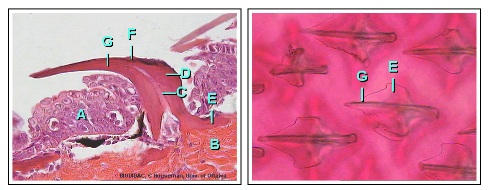
Shark teeth - Alexia Sioda
editMain Article: Shark tooth[95]
A shark tooth is one of the numerous teeth of a shark. Shark teeth are a very fascinating study subject because they are highly mineralized tissues that are continually being produced by sharks.[96] Sharks have a constant shedding of their teeth and for example, Carcharhiniformes shed approximately 35,000 teeth in a lifetime, replacing those that fall out. Several studies suggest that sharks are able to replace their teeth once a week because they grow in the gum tissue unlike most vertebrates that grow their teeth in alveoli.[97] They are only shed once new teeth are formed underneath and push them out of the connective tissue that was holding them in place.[97] There are four basic types of shark teeth: dense flattened, needle-like, pointed lower with triangular upper, and non-functional. The type of tooth that a shark has depends on its diet and feeding habits.
Fossilized evidence of teeth have dated back to almost 450 million years ago indicating the most ancient types of sharks occurred during the Late Ordovician period.[97]
The sex of the shark also plays a role in the development of teeth and the differences in teeth in species due to gender is called sexual heterodonty.[98] Usually, females have larger teeth because on average they are usually larger than males.[98] Also, age can change the shape of teeth in which "juvenile teeth start out more narrow ad robust, while adult teeth are broader and thinner".[98]
Week 5: Group Work
editTaylor Stokes
edit- Topic #1: The 5 synapomorphies that sharks evolved from.
- Articles: caudal fin[24], dorsal fin[25], gills[20], notochord[99], dorsal nerve cord[100], and pharyngeal slit[101].
- Images or other media: Endostyle[102]
- Bibliography: homology review [103]
- Topic #2: Shark organs
- Articles: Shark[104], osmoregulation[105], shark liver oil[106]
- Images or other media: anatomy
- This Image depicts the anatomy of a shark
- Bibliography: Shark liver oil[107]
Alyssa Jordan
edit- Topics with articles:
- 1) Electroreceptor organs (near nose, eyes, and mouth)
- 2) Tonic Immobility (when sharks become immobile when flipped from their belly onto their back)
- Apparent death#Tonic immobility[114]
- The tonic immobility reaction of the domestic fowl: a review[115]
- 3) Muscles
- 4) Rearrangement of "Integument" section through adding "Skin" as a subcomponent, along with ampullae of Lorenzini and placoid scales
- Images/media:
- ^ Stach, Thomas (January 1, 2002). "Minireview: On the Homology of the Protocoel in Cephalochordata and 'Lower' Deuterostomia". Acta Zoologica. 83: 25–31 – via Ebsco.
- ^ Satoh, Noriyuki (2016). "Dorsal Nerve Cord". Science Direct.
- ^ Holland, Linda (December 19, 2015). "The origin and evolution of chordate nervous systems". Philosophical transactions of the Royal Society of London. 370: 1684.
- ^ a b Kardong, Kenneth (2019). Vertebrates: Comparative Anatomy, Function, Evolution. McGraw-Hill. pp. 51–56.
- ^ Kardong, Kenneth (2019). Vertebrates: Comparative Anatomy, Evolution, Function. McGraw-Hill. pp. 80–86.
- ^ Holland, Linda (December 19, 2015). "The origin and evolution of chordate nervous systems". Philosophical transactions of the Royal Society of London. 370: 1–8.
- ^ Risbud, Makarand (January 21, 2011). "Notochordal cells in the adult intervertebral disc: new perspective on an old question". Crit Rev Eukaryot Gene Expr. 1: 29–41.
- ^ Awruch, Cynthia (November 17, 2015). "Reproduction Strategies". Science Direct. 34: 255–310.
- ^ Argyriou, Thodoris (Jan 6, 2016). "Exceptional preservation reveals gastrointestinal anatomy and evolution in early actinopterygian fishes". Science reports. 6.
- ^ Piermarini, Peter (January 2004). "Homeostasis: osmoregulation, pH regulation, and nitrogen excretion". Research Gate: 247–268.
- ^ "How Does the Oily Liver of a Shark Work?". Pets on Mom.com. Retrieved 2021-03-27.
- ^ Reilly, Beau (September 1, 2011). "Branchial osmoregulation in the euryhaline bull shark, Carcharhinus leucas: a molecular analysis of ion transporters". The Company of Biologists. 17: 2883–2895.
- ^ Hasegawa, Kumi (April 27, 2016). "Sulfate transporters involved in sulfate secretion in the kidney are localized in the renal proximal tubule II of the elephant fish (Callorhinchus milii)". American Journal of Physiology. 1: 66–78.
- ^ Stephenson, Andrea (December 2016). "The vertebrate heart: an evolutionary perspective". Journal of Anatomy. 6: 787–797.
- ^ "Shark anatomy 101". CHINCOTEAGUE BAY FIELD STATION. Retrieved 2021-03-27.
- ^ "Dogfish Internal Anatomy". www.zoology.ubc.ca. Retrieved 2021-03-27.
- ^ Martin, Aidan. "The Importance of Being Cartilaginous". ReefQuest Centre for Shark Research.
- ^ "Biomimicry Shark Denticles | Smithsonian Ocean". ocean.si.edu. Retrieved 2020-12-09.
- ^ "Muscle movement anatomy of the great white shark". ultimate-animals.com. Retrieved 2020-12-09.
- ^ a b c d Kardong, Kenneth V. (2019). Vertebrates: comparative anatomy, function, evolution. New York: McGraw-Hill Education. pp. 213–217. ISBN 978-1-259-70091-0. Cite error: The named reference ":3" was defined multiple times with different content (see the help page).
- ^ a b c d e "Fish scale", Wikipedia, 2021-02-01, retrieved 2021-03-27
- ^ Britannica, The Editors of Encyclopaedia. "Scale". Encyclopedia Britannica, 7 Jul. 2011, https://www.britannica.com/science/scale-zoology. Accessed 26 March 2021.
- ^ "Electroreception", Wikipedia, 2021-02-23, retrieved 2021-03-27
- ^ a b c d e f g "Fish fin", Wikipedia, 2021-03-08, retrieved 2021-03-12
- ^ a b c d e f g "Dorsal fin", Wikipedia, 2020-12-05, retrieved 2021-03-12
- ^ "Shark Anatomy". Shark Trust. 2020. Retrieved March 26, 2020.
{{cite web}}: CS1 maint: url-status (link) - ^ Martin, Aidan. "The Importance of Being Cartilaginous". ReefQuest Centre for Shark Research.
- ^ "Biomimicry Shark Denticles | Smithsonian Ocean". ocean.si.edu. Retrieved 2020-12-09.
- ^ "Muscle movement anatomy of the great white shark". ultimate-animals.com. Retrieved 2020-12-09.
- ^ a b c Kardong, Kenneth V. (2019). Vertebrates: comparative anatomy, function, evolution. New York: McGraw-Hill Education. pp. 213–217. ISBN 978-1-259-70091-0.
- ^ Britannica, The Editors of Encyclopaedia. "Scale". Encyclopedia Britannica, 7 Jul. 2011, https://www.britannica.com/science/scale-zoology. Accessed 26 March 2021.
- ^ "Electroreception", Wikipedia, 2021-02-23, retrieved 2021-03-27
- ^ "Shark Anatomy". Shark Trust. 2020. Retrieved March 26, 2020.
{{cite web}}: CS1 maint: url-status (link) - ^ Stach, Thomas (January 1, 2002). "Minireview: On the Homology of the Protocoel in Cephalochordata and 'Lower' Deuterostomia". Acta Zoologica. 83: 25–31 – via Ebsco.
- ^ "lateral - Wiktionary". en.wiktionary.org. Retrieved 2021-03-27.
- ^ Holland, Linda (December 19, 2015). "The origin and evolution of chordate nervous systems". Philosophical transactions of the Royal Society of London. 370: 1684.
- ^ a b Kardong, Kenneth (2019). Vertebrates: Comparative Anatomy, Function, Evolution. McGraw-Hill. pp. 51–56.
- ^ Kardong, Kenneth (2019). Vertebrates: Comparative Anatomy, Evolution, Function. McGraw-Hill. pp. 80–86.
- ^ Awruch, Cynthia (November 17, 2015). "Reproduction Strategies". Science Direct. 34: 255–310.
- ^ Argyriou, Thodoris (Jan 6, 2016). "Exceptional preservation reveals gastrointestinal anatomy and evolution in early actinopterygian fishes". Science reports. 6.
- ^ Piermarini, Peter (January 2004). "Homeostasis: osmoregulation, pH regulation, and nitrogen excretion". Research Gate: 247–268.
- ^ "How Does the Oily Liver of a Shark Work?". Pets on Mom.com. Retrieved 2021-03-27.
- ^ Reilly, Beau (September 1, 2011). "Branchial osmoregulation in the euryhaline bull shark, Carcharhinus leucas: a molecular analysis of ion transporters". The Company of Biologists. 17: 2883–2895.
- ^ Hasegawa, Kumi (April 27, 2016). "Sulfate transporters involved in sulfate secretion in the kidney are localized in the renal proximal tubule II of the elephant fish (Callorhinchus milii)". American Journal of Physiology. 1: 66–78.
- ^ Stephenson, Andrea (December 2016). "The vertebrate heart: an evolutionary perspective". Journal of Anatomy. 6: 787–797.
- ^ "Shark anatomy 101". CHINCOTEAGUE BAY FIELD STATION. Retrieved 2021-03-27.
- ^ "Dogfish Internal Anatomy". www.zoology.ubc.ca. Retrieved 2021-03-27.
- ^ Stach, Thomas (January 1, 2002). "Minireview: On the Homology of the Protocoel in Cephalochordata and 'Lower' Deuterostomia". Acta Zoologica. 83: 25–31 – via Ebsco.
- ^ "lateral - Wiktionary". en.wiktionary.org. Retrieved 2021-03-27.
- ^ a b Kardong, Kenneth (2019). Vertebrates: Comparative Anatomy, Function, Evolution. McGraw-Hill. pp. 51–56.
- ^ Kardong, Kenneth (2019). Vertebrates: Comparative Anatomy, Evolution, Function. McGraw-Hill. pp. 80–86.
- ^ "How Does the Oily Liver of a Shark Work?". Pets on Mom.com. Retrieved 2021-03-27.
- ^ "Shark anatomy 101". CHINCOTEAGUE BAY FIELD STATION. Retrieved 2021-03-27.
- ^ "Dogfish Internal Anatomy". www.zoology.ubc.ca. Retrieved 2021-03-27.
- ^ Martin, Aidan. "The Importance of Being Cartilaginous". ReefQuest Centre for Shark Research.
- ^ "Biomimicry Shark Denticles | Smithsonian Ocean". ocean.si.edu. Retrieved 2020-12-09.
- ^ "Muscle movement anatomy of the great white shark". ultimate-animals.com. Retrieved 2020-12-09.
- ^ Britannica, The Editors of Encyclopaedia. "Scale". Encyclopedia Britannica, 7 Jul. 2011, https://www.britannica.com/science/scale-zoology. Accessed 26 March 2021.
- ^ "Electroreception", Wikipedia, 2021-02-23, retrieved 2021-03-27
- ^ "Shark Anatomy". Shark Trust. 2020. Retrieved March 26, 2020.
{{cite web}}: CS1 maint: url-status (link) - ^ "Biomimicry Shark Denticles | Smithsonian Ocean". ocean.si.edu. Retrieved 2020-12-09.
- ^ "Muscle movement anatomy of the great white shark". ultimate-animals.com. Retrieved 2020-12-09.
- ^ "Muscle movement anatomy of the great white shark". ultimate-animals.com. Retrieved 2020-12-09.
- ^ "Chondrichthyes", Wikipedia, 2021-03-14, retrieved 2021-03-26
- ^ "Ampullae of Lorenzini", Wikipedia, 2020-12-29, retrieved 2021-03-16
- ^ "Ichthyology", Wikipedia, 2021-02-04, retrieved 2021-03-26
- ^ "Stefano Lorenzini", Wikipedia, 2021-01-10, retrieved 2021-03-26
- ^ "Electromagnetic field", Wikipedia, 2021-02-21, retrieved 2021-03-27
- ^ "Passive electrolocation in fish", Wikipedia, 2020-08-30, retrieved 2021-03-27
- ^ "Electroreception", Wikipedia, 2021-02-23, retrieved 2021-03-27
- ^ Britannica, The Editors of Encyclopaedia. "Scale". Encyclopedia Britannica, 7 Jul. 2011, https://www.britannica.com/science/scale-zoology. Accessed 26 March 2021.
- ^ "Salmon shark", Wikipedia, 2021-03-07, retrieved 2021-03-27
- ^ "Tuna", Wikipedia, 2021-03-08, retrieved 2021-03-27
- ^ "Shark Anatomy". Shark Trust. 2020. Retrieved March 26, 2020.
{{cite web}}: CS1 maint: url-status (link) - ^ a b c "Pharyngeal slit", Wikipedia, 2021-03-10, retrieved 2021-03-16
- ^ a b c d "Dorsal nerve cord", Wikipedia, 2020-12-17, retrieved 2021-03-16
- ^ a b c "Notochord", Wikipedia, 2021-01-28, retrieved 2021-03-16
- ^ a b c "Endostyle", Wikipedia, 2021-03-16, retrieved 2021-03-16
- ^ "lateral - Wiktionary". en.wiktionary.org. Retrieved 2021-03-27.
- ^ "Gill", Wikipedia, 2021-02-07, retrieved 2021-03-12
- ^ Stach, Thomas (January 1, 2002). "Minireview: On the Homology of the Protocoel in Cephalochordata and 'Lower' Deuterostomia". Acta Zoologica. 83: 25–31 – via Ebsco.
- ^ "Shark", Wikipedia, 2021-03-01, retrieved 2021-03-16
- ^ "Clasper", Wikipedia, 2021-03-14, retrieved 2021-03-27
- ^ "Oviduct", Wikipedia, 2020-11-27, retrieved 2021-03-27
- ^ "Shark", Wikipedia, 2021-03-01, retrieved 2021-03-27
- ^ "Shark", Wikipedia, 2021-03-01, retrieved 2021-03-27
- ^ "Shark liver oil", Wikipedia, 2021-03-10, retrieved 2021-03-16
- ^ "How Does the Oily Liver of a Shark Work?". Pets on Mom.com. Retrieved 2021-03-27.
- ^ "Osmoregulation", Wikipedia, 2021-02-28, retrieved 2021-03-16
- ^ "Shark", Wikipedia, 2021-03-01, retrieved 2021-03-27
- ^ "Shark", Wikipedia, 2021-03-01, retrieved 2021-03-27
- ^ "Shark", Wikipedia, 2021-03-01, retrieved 2021-03-27
- ^ "Shark anatomy 101". CHINCOTEAGUE BAY FIELD STATION. Retrieved 2021-03-27.
- ^ "Dogfish Internal Anatomy". www.zoology.ubc.ca. Retrieved 2021-03-27.
- ^ "Shark tooth", Wikipedia, 2021-03-01, retrieved 2021-03-16
- ^ Enax, Joachim (June 2012). "Structure, composition, and mechanical properties of shark teeth". Journal of Structural Biology. 178: 290–299 – via Elsevier Science Direct.
- ^ a b c "Ancient Sharks". Micronesian Conservation Coalition. 2015.
{{cite web}}: CS1 maint: url-status (link) - ^ a b c Fossil Shark Teeth. The Paleontological Society. pp. 1–2.
- ^ "Notochord", Wikipedia, 2021-01-28, retrieved 2021-03-16
- ^ "Dorsal nerve cord", Wikipedia, 2020-12-17, retrieved 2021-03-16
- ^ "Pharyngeal slit", Wikipedia, 2021-03-10, retrieved 2021-03-16
- ^ "Endostyle", Wikipedia, 2021-03-16, retrieved 2021-03-16
- ^ Stach, Thomas (January 1, 2002). "Minireview: On the Homology of the Protocoel in Cephalochordata and 'Lower' Deuterostomia". Acta Zoologica. 83: 25–31 – via Ebsco.
- ^ "Shark", Wikipedia, 2021-03-01, retrieved 2021-03-16
- ^ "Osmoregulation", Wikipedia, 2021-02-28, retrieved 2021-03-16
- ^ "Shark liver oil", Wikipedia, 2021-03-10, retrieved 2021-03-16
- ^ Samimi, Nastaran (October 2020). "The Therapeutic Effect of Shark Liver Oil in a Rat Model of Acetic Acid-Induced Ulcerative Colitis". Evidence-Based Complementary & Alternative Medicine (ECAM): 1–8 – via Ebsco.
- ^ "Electroreception", Wikipedia, 2021-02-23, retrieved 2021-03-16
- ^ "Electric fish", Wikipedia, 2021-03-02, retrieved 2021-03-16
- ^ "Passive electrolocation in fish", Wikipedia, 2020-08-30, retrieved 2021-03-16
- ^ "Ampullae of Lorenzini", Wikipedia, 2020-12-29, retrieved 2021-03-16
- ^ Freitas, Renata (January 2006). "Developmental Origin of Shark Electrosensory Organs". Evolution & Development. 8: 74–80 – via EBSCOhost.
- ^ Modrell, Melinda (May 2012). "Evolution of Electrosensory Ampullary Organs: Conservation of Eya4 Expression during Lateral Line Development in Jawed Vertebrates". Evolution & Development. 14: 277–285 – via EBSCOhost.
- ^ "Apparent death", Wikipedia, 2021-01-28, retrieved 2021-03-16
- ^ Bryan, Jones (1986). "The tonic immobility reaction of the domestic fowl: a review". World's Poultry Science Journal. 42: 82–96 – via Taylor&FrancisOnline.
- ^ Bernal, Diego (October 2005). "Mammal-like Muscles Power Swimming in a Cold-Water Shark". Nature. 437: 1349–1352 – via EBSCOhost.
- ^ Mallatt, Jon (October 1997). "Shark Pharyngeal Muscles and Early Vertebrate Evolution". Acta Zoologica. 78: 279–94 – via EBSCOhost.
- ^ Chris_huh (2007-03-06), Electroreceptors in a sharks head, including Ampullae of Lorenzini and Lateral Line canals, retrieved 2021-03-16
Alexia Sioda
edit- Topics: shark skin function and shark teeth
- Articles: Denticle[1], Shark tooth[2], Hyaline cartilage[3], Poikilotherm[4]
- Images or other media:
- References:
Feedback: Discussion and Adding to an Article
edit- I love how you are all drafting in your own sandbox first, using links AND adding citations and then adding to the group sandbox. This has put you in excellent position to develop a really good draft.
- I am also delighted to see that I can easily tell who is working on what so contribution tracking is easy.
- This week I want to REALLY REALLY encourage you to submit proposed changes you are thinking about to the talk pages. This is a really helpful practice.
- Your objective for next week is to create a working draft and you are already well on your way with the hard work you have already put in. I encourage you to continue cross-checking for other articles that may be covering the topics you are all interested in...but really, you are doing great.
- now is the time to think about where you are feeling unsure and what you want my feedback on. Are you afraid that you took on too much? Do you need to cut back? Should you prioritize your edits?Osquaesitor (talk) 21:19, 23 March 2021 (UTC)
- ^ "Denticle", Wikipedia, 2019-12-15, retrieved 2021-03-16
- ^ "Shark tooth", Wikipedia, 2021-03-01, retrieved 2021-03-16
- ^ "Hyaline cartilage", Wikipedia, 2021-01-17, retrieved 2021-03-16
- ^ "Poikilotherm", Wikipedia, 2021-02-24, retrieved 2021-03-16
- ^ Domel, August (August 2, 2018). "Hydrodynamic properties of biomimetic shark skin: effect of denticle size and swimming speed" (PDF). Bioinspir. Biomim. 13: 1–15 – via IOP Publishing.
- ^ Enax, Joachim (June 2012). "Structure, composition, and mechanical properties of shark teeth". Journal of Structural Biology. 178: 290–299 – via Elsevier Science Direct.