== Anatomy of the heart == BY Tariq aqel
The heart is a hollow muscular organ of a somewhat conical form; it lies between the lungs in the middle mediastinum and is enclosed in the pericardium . It is placed obliquely in the chest behind the body of the sternum and adjoining parts of the rib cartilages, and projects farther into the left than into the right half of the thoracic cavity, so that about one-third of it is situated on the right and two-thirds on the left of the median plane.
Size.—The heart, in the adult, measures about 12 cm. in length, 8 to 9 cm. in breadth at the broadest part, and 6 cm. in thickness. Its weight, in the male, varies from 280 to 340 grams; in the female, from 230 to 280 grams. The heart continues to increase in weight and size up to an advanced period of life; this increase is more marked in men than in women.
Component Parts the heart is subdivided by septa into right and left halves, and a constriction subdivides each half of the organ into two cavities, the upper cavity being called the atrium, the lower the ventricle. The heart therefore consists of four chambers, viz., right and left atria, and right and left ventricles.
The division of the heart into four cavities is indicated on its surface by grooves. The atria are separated from the ventricles by the coronary sulcus (auriculoventricular groove); this contains the trunks of the nutrient vessels of the heart, and is deficient in front, where it is crossed by the root of the pulmonary artery. The interatrial groove, separating the two atria, is scarcely marked on the posterior surface, while anteriorly it is hidden by the pulmonary artery and aorta. The ventricles are separated by two grooves, one of which, the anterior longitudinal sulcus, is situated on the sternocostal surface of the heart, close to its left margin, the other posterior longitudinal sulcus, on the diaphragmatic surface near the right margin; these grooves extend from the base of the ventricular portion to a notch, the incisura apicis cordis, on the acute margin of the heart just to the right of the apex.
The Right Side of Your Heart
In figure ,the superior and inferior vena cavae are shown in blue to the left of the heart muscle as you look at the picture. These veins are the largest veins in your body. After your body's organs and tissues have used the oxygen in your blood, the vena cavae carry the oxygen-poor blood back to the right atrium of your heart.
The superior vena cava carries oxygen-poor blood from the upper parts of your body, including your head, chest, arms, and neck. The inferior vena cava carries oxygen-poor blood from the lower parts of your body.
The oxygen-poor blood from the vena cavae flows into your heart's right atrium and then to the right ventricle. From the right ventricle, the blood is pumped through the pulmonary (PULL-mun-ary) arteries (shown in blue in the center of figures) to your lungs.
Once in the lungs, the blood travels through many small, thin blood vessels called capillaries. There, the blood picks up more oxygen and transfers carbon dioxide to the lungs—a process called gas exchange' The oxygen-rich blood passes from your lungs back to your heart through the pulmonary veins.
The Left Side of Your Heart
Oxygen-rich blood from your lungs passes through the pulmonary veins (shown in red to the right of the left atrium in figures). The blood enters the left atrium and is pumped into the left ventricle.
From the left ventricle, the oxygen-rich blood is pumped to the rest of your body through the aorta. The aorta is the main artery that carries oxygen-rich blood to your body.
Like all of your organs, your heart needs oxygen-rich blood. As blood is pumped out of your heart's left ventricle, some of it flows into the coronary arteries (shown in red in figure below).
Your coronary arteries are located on your heart's surface at the beginning of the aorta. They carry oxygen-rich blood to all parts of your heart.
The heart is demarcated by:
-A point 9 cm to the left of the midsternal line (apex of the heart)
-The seventh right sternocostal articulation
-The upper border of the third right costal cartilage 1 cm from the right sternal line
-The lower border of the second left costal cartilage 2.5 cm from the left lateral sternal line.
Cardiac Conduction
| طارق عقل1993 | |
|---|---|
 Isolated conduction system of the heart | |
 Heart; conduction system | |
| Details | |
| Identifiers | |
| Latin | systema conducente cordis |
| Anatomical terminology | |
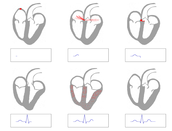
Cardiac conduction is the rate at which the heart conducts electrical impulses. Cardiac muscle cells contract spontaneously. These contractions are coordinated by the sinoatrial (SA) node which is also referred to as the pacemaker of the heart. The SA node is composed of nodal tissue that has characteristics of both muscle and nervous tissue. The SA node is located in the upper wall of the right atrium. When the SA node contracts it generates nerve impulses that travel throughout the heart wall causing both atria to contract.
Another section of nodal tissue lies on the right side of the partition that divides the atria, near the bottom of the right atrium. It is called the atrioventricular (AV) node. When the impulses reach the AV node they are delayed for about a tenth of a second. This delay allows the atria to contract and empty their contents first. The impulses are then sent down the atrioventricular bundle. This bundle of fibers branches off into two bundles and the impulses are carried down the center of the heart to the left and right ventricles.
At the base of the heart the atrioventricular bundles start to divide further into Purkinje fibers. When the impulses reach these fibers they trigger the muscle fibers in the ventricles to contract.

heart attack
The heart works 24 hours a day, pumping oxygen and nutrient-rich blood to the body. Blood is supplied to the heart through its coronary arteries. If a blood clot suddenly blocks a coronary artery, it cuts off most or all blood supply to the heart, and a heart attack results. Cells in the heart muscle that do not receive enough oxygen-rich blood begin to die. The more time that passes without treatment to restore blood flow, the greater the damage to the heart. Having a heart attack is a big wakeup call for most people. There's usually no problem convincing a heart attack survivor to change his or her lifestyle-to quit smoking, control their high blood pressure and cholesterol, control their weight and get regular physical activity.
Each year, more than one million people in the U.S. have a heart attack and about half - 515,000 - of them die. Half of those who die do so within one hour of the start of symptoms and before reaching the hospital.
A heart attack is an emergency. Call emergency services if you think you or someone else may be having a heart attack. Prompt treatment of a heart attack can help prevent or limit damage to the heart and prevent sudden death.
It is important to call emergency services fast - within 5 minutes - because emergency personnel can give a variety of treatments and medicines at the scene. They carry drugs and equipment that can help your medical condition, including
- oxygen
- aspirin to prevent further blood clotting
- heart medications, such as nitroglycerin
- pain relief treatments
- defibrillators that can restart the heart if it stops beating.
If blood flow in the blocked artery can be restored quickly, permanent heart damage may be prevented. Yet, many people do not seek medical care for 2 hours or more after symptoms start. The symptoms of a heart attack can include chest discomfort, discomfort in other areas of the upper body, shortness of breath, and other symptoms.
The most common symptom of heart attack is chest pain or discomfort. Most heart attacks involve discomfort in the center of the chest that lasts for more than a few minutes or goes away and comes back. The discomfort can feel like uncomfortable pressure, squeezing, fullness, or pain. It can be mild or severe. Heart attack pain can sometimes feel like indigestion or heartburn.
Discomfort can also occur in other areas of the upper body, including pain or numbness in one or both arms, the back, neck, jaw or stomach. Shortness of breath often happens along with, or before chest discomfort. Other symptoms may include breaking out in a cold sweat, having nausea and vomiting, or feeling light-headed or dizzy or fainting.
Signs and symptoms vary from person to person. In fact, if you have a second heart attack, your symptoms may not be the same as the first heart attack. Some people have no symptoms. This is called a "silent" heart attack.
Angina is chest pain or discomfort that occurs when your heart muscle does not get enough blood. Angina symptoms can be very similar to heart attack symptoms. If you have angina and notice a sudden change or worsening of your symptoms, talk with your doctor right away.
