Talk:Phase-contrast microscopy
| This article is rated Start-class on Wikipedia's content assessment scale. It is of interest to the following WikiProjects: | |||||||||||||||||||||
| |||||||||||||||||||||
.jpg)
.jpg)
Wrong direction of phase or swapped positive/negative phase imaging?
edit- First, the background light is phase-shifted by −90° by passing it through a phase-shift ring, which eliminates the phase difference between the background and the scattered light rays
As seen from what is written later, in negative phase contrast this leads to -90 + (-90) = -180 degrees phase retardation, whereas in positive phase contrast one would eliminate the phase difference 45.141.152.52 (talk) 19:11, 7 November 2019 (UTC)
Phase shift misleading
editIn the section 'technical details', the following sentence:
- Assuming that the specimen does not significantly alter the amplitudes of the incoming wavefronts but mainly changes phase relations with respect to the "unperturbed" wavefronts, newly generated spherical wave fronts that are retarded by 90° (λ/4) emanate from 'O'
Is wrong/misleading. The wave fronts need not be shifted by exactly 90°. The shift is equal to the difference in the optical path length (which depends on the thickness and refractive index of the sample) divided by the wavelength of the light. In fact, if it were always exactly 90°, it wouldn't be able to resolve a picture - the destructive interference would result in a uniformly black image.
Granted, most practical phase contrast setups are designed so the phase shift is around 90°. However, this is only an average, and for it to be true we must take into account the typical thicknesses of biological specimens, the refractive index of water, and the wavelengths of light that are used. — Preceding unsigned comment added by 130.216.209.138 (talk) 02:51, 5 December 2011 (UTC)
Explanation seems incomplete/inconsistent
editThe Related Methods states that phase-contrast microscopy uses polarized light, yet the Working Principle section does not mention the light being polarized. It also seems to me that true phase-shift detection would require a coherent light source so that scattered and background photons would start in a known phase relationship. Also, there is no mention of how this is an improvement on dark-field microscopy, where the background is black and all you see is the scattered light. Finally, in the Related Methods it is mentioned that phase-contrast microscopy is "unsuitable where the object ... alter(s) polarization" yet that is the entire point of the phase-contrast system: to see the object through its effect on polarization. Perhaps that text should read "unsuitable where the container for the object alters polarization."198.91.146.14 (talk) 04:14, 19 February 2019 (UTC)
Merger
editI agree that they should be merged.James.folsom 21:34, 16 January 2007 (UTC)
After considering the circumstances behind editing articles in Wikipedia (and keeping them free of commercial links!), I propose having this article be merged into "phase contrast microscope". Gregor
- OK, merger is taken care of. Please see Talk:Phase_contrast_microscope for fuller discussion of the merge, and earlier discussion. Peter G Werner (talk) 23:55, 15 February 2008 (UTC)
Refspam?
editThese look like WP:REFSPAM. Are they all actually verifying unique content? If so, I apologise. Let's look. --Ronz (talk) 18:31, 3 October 2014 (UTC)
They were all added as inline citations with this rewrite, so maybe they are indeed verifying something, but were all just thrown at the end instead of where they actually verify unique content. --Ronz (talk) 18:58, 3 October 2014 (UTC)
Seems that we should have better sources than those of two manufacturers, one of which is a dead link. --Ronz (talk) 19:08, 3 October 2014 (UTC)
External links
editWhile cleaning up after Amadori Heyns (talk · contribs)'s spamming of leica-microsystems.com, I came across this article. I removed the external links section because the links appeared to fail WP:EL, especially WP:ELNO#1. I've copied them below for discussion. --Ronz (talk) 18:42, 3 October 2014 (UTC)
- Historic phase contrast microscopy movies made by Kurt Michel at Carl Zeiss AG in the early 1940s
- I don't see how these help in the understanding of the topic of the article, given we've already Figure 1. --Ronz (talk) 18:50, 3 October 2014 (UTC)
- Optical Microscopy Primer — Phase Contrast Microscopy by Florida State University
- Given the extent of information and resources, it might be worth keeping. --Ronz (talk) 18:50, 3 October 2014 (UTC)
- Differential Interference Contrast (DIC) Microscopy by Nikon
- Too far off topic to begin, and too little directly on DIC besides just providing a directory of resources. --Ronz (talk) 18:50, 3 October 2014 (UTC)
- Hoffman Modulation Contrast Basics by Olympus
- Off topic. --Ronz (talk) 18:52, 3 October 2014 (UTC)
- Quantitative Phase Contrast Microscopy by Phase Holographic Imaging
- Off topic. --Ronz (talk) 18:53, 3 October 2014 (UTC)
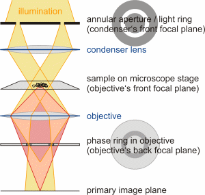
Ray path image is wrong.
editI believe the image used to illustrate the light cones is wrong.
This is the image currently being used:
As you can see, the impression is given that the light from the condensor annulus is focused onto a point of the specimen.
This is a different image describing the process:
https://home.uni-leipzig.de/pwm/web/img/intro_phasecontrast_2.png
Here, one can see that the annulus is not point focused onto the specimen, but illuminates it broadly.
This makes much more sense, because then the background light does not suffer the exact same phase shift in the specimen as the specimen-scattered light - in the image as it stand presently, no phase shift would occur!
I am awaiting comments, and will then delete the erroneous image. I am not sure if we can use the other image, as it seems copyrighted. 00:30, 11 April 2020 (UTC)2A02:8109:B540:7CB:DD32:A67F:10F3:768F (talk)
This page is of exceptionally poor quality
editThis page does a pretty poor job of explaining phase contrast and irrelevant references to other imaging techniques like DIC of quantitative phase contrast (advertisement?). Claims that Hofman modulation contrast is still popular are laughable. Show would you guys feel about a bigger rewrite? --Frozenport (talk) 21:41, 17 April 2020 (UTC)