| This is the talk page for discussing improvements to the Parietal eye article. This is not a forum for general discussion of the article's subject. |
Article policies
|
| Find sources: Google (books · news · scholar · free images · WP refs) · FENS · JSTOR · TWL |
| This article is rated Start-class on Wikipedia's content assessment scale. It is of interest to the following WikiProjects: | |||||||||||||||||||||
| |||||||||||||||||||||

This article may be too technical for most readers to understand. (September 2010) |
|
This article links to one or more target anchors that no longer exist.
Please help fix the broken anchors. You can remove this template after fixing the problems. | Reporting errors |
Where is it?
editThe article doesn't say where exactly on the tuatara this third eye is located. The picture may suggest something but isn't entirely clear either. Thanks. -- 71.71.192.231 (talk) 01:51, 22 December 2007 (UTC)
- I agree. Could someone perhaps modify the image with an arrow or similar marking? It is not very clear. Thanks. -- 125.236.161.221 (talk) 02:58, 5 January 2008 (UTC)
- the text says on the forehead, especially of the young. the image shows an adult in side view. does anyone just have a different image? of a young one in front top view? —Preceding unsigned comment added by 208.54.94.60 (talk) 13:27, 13 March 2008 (UTC)
The language is almost entirely technical and inaccessible to a general reader, it needs to use words like "head" "mounth" "between the eyes" but I can't even figure out where to start amongst all the epi-whatevers.~~
I added a new image (28 August) of an iguana with a much clearer parietal eye. Hope this helps! SurreyJohn (Talk) 11:55, 30 October 2014 (UTC)
Physiology
editI thought the section on Physiology was vague and un-descriptive. I wrote an alternative, but I want to submit it for concurrence in case I made a mistake. I also need more references about the structure in other classes like those of fish.
The parietal eye is a part of the pineal complex, which is a part of the epithalamus located on the diencephalon. The term is used to describe multiple specific structures in different species. The parietal eye can be a frontal (parapineal) organ which either penetrates the skull (such as in frogs) or does not penetrate it (salamanders). In other animals, the parietal eye is a part of the epiphysis (the pineal organ, or pineal gland if mostly endocrine), such as with lizards, tuatara, and fish, where it may resemble the parietal organ by being an anterior evagination. In most cases it contains photoreceptive cells but may not be capable of receiving light.[1] The parietal eye uses a different biochemical method of detecting light than rod cells or cone cells in a normal vertebrate eye.[2]
Here is the original for comparison.
The parietal eye is a part of the epithalamus, which can be divided into two major parts; the epiphysis (the pineal organ, or pineal gland if mostly endocrine) and the parietal organ (often called the parietal eye, or third eye if it is photoreceptive). It arises as an anterior evagination of the pineal organ or as a separate outgrowth of the roof of the diencephalon. In some species, it protrudes through the skull.[3] The parietal eye uses a different biochemical method of detecting light than rod cells or cone cells in a normal vertebrate eye.[4]
This first sentence in the Anatomy section is fallacious: It says that the "parietal eye" is subdivided into pineal gland and "parietal eye".... I think this should be clarified... — Preceding unsigned comment added by 132.230.96.138 (talk) 09:22, 4 November 2016 (UTC)
- The Tuatara article states that the Tuatara's parietal eye "has its own lens, cornea, retina with rod-like structures, and degenerated nerve connection to the brain, suggesting it evolved from a real eye." The phrase "rod-like structures" appears to conflict with both the original and re-written version, and should be clarified at some point by someone who is familiar with the Tuatara's rod-like structures. —Preceding unsigned comment added by 72.244.206.65 (talk) 22:30, 15 May 2011 (UTC)
References
- ^ Zug, George (2002). Herpetology: An Introductory Biology of Amphibians and Reptiles, Second Edition. San Diego: Academic Press. p. 75. ISBN 0-12-782622-X.
{{cite book}}: Unknown parameter|coauthors=ignored (|author=suggested) (help) - ^ Xiong, Wei-Hong (1998). "An unusual cGMP pathway underlying depolarizing light response of the vertebrate parietal-eye photoreceptor". Nature Neuroscience. 1: 359–65. doi:10.1038/1570. Retrieved 2007-02-22.
{{cite journal}}: Unknown parameter|coauthors=ignored (|author=suggested) (help) - ^ Zug, George (2002). Herpetology: An Introductory Biology of Amphibians and Reptiles, Second Edition. San Diego: Academic Press. p. 75. ISBN 0-12-782622-X.
{{cite book}}: Unknown parameter|coauthors=ignored (|author=suggested) (help) - ^ Xiong, Wei-Hong (1998). "An unusual cGMP pathway underlying depolarizing light response of the vertebrate parietal-eye photoreceptor". Nature Neuroscience. 1: 359–65. doi:10.1038/1570. Retrieved 2007-02-22.
{{cite journal}}: Unknown parameter|coauthors=ignored (|author=suggested) (help)
Function
editThe function section merely goes over morphology in various groups, it says nothing about the actual function of the structure. Troodon311 (talk) 14:21, 26 May 2010 (UTC)
- Indeed. And I'm too lazy to do anything about it. Just like everyone else. —iNkubusse? 11:28, 30 January 2012 (UTC)
- Man I was really disappointed too, I was really hoping to learn what the 3rd eye did but it just listed unrelated stuff.129.2.129.218 (talk) 07:17, 1 February 2013 (UTC)Mason
Bad reference
editThis reference:
^ "Parietal eye". Tuatara Glossary. School of Biological Sciences, Victoria University of Wellington. 2007-09-11. Retrieved 2008-05-28.
Does not refer to the parietal eye, and therefore should be removed. I tried removing it, but it was reverted. Rhetth (talk) 17:18, 10 January 2011 (UTC)
- Done. I looked for a replacement link, but could not find it. Luckily the actual quotation comes from a different site, so no info had to be removed. --TheAlphaWolf (talk) 13:49, 11 January 2011 (UTC)
- A replacement link was easy to find at the Internet Archive's Wayback Machine. --Stemonitis (talk) 14:12, 11 January 2011 (UTC)
Presence in various animals?
editThis section has problems. First it starts with sensationalistic material about tuatara instead of just explaining that this organ is common in ectothermic (cold-blooded) vertebrates. The part about a "poorly developed" version in salamanders makes no sense, since there are hundreds of species. Some frogs and salamanders can detect linearly polarized light from the sky and navigate using their parietal eyes. Zyxwv99 (talk) 21:25, 14 November 2015 (UTC)
Iguana Parietal eye.JPG Image
edit
I have moved the following question/discussion after User:Stas000D deleted this file. Discusses of articles and content should be done on the relevant article's talk page so anyone can see and join in. SJ.
Hello. Why do you think that every spot on a lizard's head is a parietal eye? Stas (talk) 20:16, 7 January 2016 (UTC)
- @Stas000D:: Clearly I do not think every spot on a lizard head is a parietal eye! However, this one does appear very distinct, it is in the correct position, top of head, central, and between the eyes. The Madagascan iguana is well known for its "third eye". See http://www.exotic-pets.co.uk/three-eyed-madagascan-iguana.html as a simple example. SurreyJohn (Talk) 23:35, 12 January 2016 (UTC)
- Dear colleague,
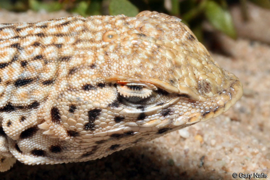
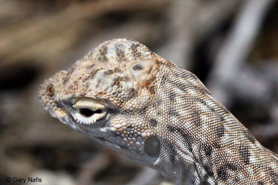
- I have written a bit about parietal eyes (with the help of more reliable sources than a shop of exotic pets) and can tell this eye from a patch of dark skin. Size of parietal eye of extant reptiles is several tenths of millimeter, rarely up to 1 mm. Eakin R. M. (1973). The Third Eye. University of California Press. pp. 1–2.: "This animal... has one of the best third eyes known." and "The third eye of S. occidentalis is barely visible to the unaided human eye...". Page 34: "The reader should be reminded of the small size of the parietal eye (about 0.2 mm in diameter)." Edinger T. (1955). "The size of parietal foramen and organ in reptiles. A rectification". Bulletin of the Museum of Comparative Zoology at Harvard College. 114 (1): 9.: "Spencer was amazed to find the axis of the eye 0.4 mm. long both in a 2-foot long Sphenodon and in a 6-foot long Varanus (1886, p. 183). Longitudinal and transverse diameters varied from 0.18 to 0.20 mm. and from 0.06 to 0.07 mm. in the Anniella specimens of Coe and Kunkel (1906, p. 393); mediodorsal skull length is about 8 mm., and length of the animals 105 to 152 mm. from snout to cloaca, plus a post-cloacal portion varying from 16 to 75 mm. (ibid., p. 351). Slightly larger parietal eye diameters in Phrynosoma have been expressed in microns: 258μ and 171μ (Ritter, 1891, p. 212). In short, the diameters of measured lizard parietal eyes were fractions of millimeters.". Also, it would be useful to familiarize yourself with outer appearance of parietal eyes in general: Cyclura, Varanus, Iguana, Uma, Callisaurus, Sceloporus 1 and Sceloporus 2. Moreover, in most pictures the eye itself is not even seen, because it is always hidden below the skin and scales, and the visible spot is just the pineal window - a small area of depigmented and translucent skin. What about Oplurus cyclurus itself and oplurines in general, we can see that they ...likely form a monophyletic group, corroborated by the possession of... and a black interparietal spot (Etheridge, 1969a)., in other words - the jet black spot that overlies the interparietal scale. Better photo of the same species shows that the real pineal window (which is not discernible on your picture at all) is just a small light-coloured point in the center of this black scale. And, of course, the parietal eye itself is not seen even there. Stas (talk) 07:38, 15 January 2016 (UTC)
- Thank you for your detailed reply. With your obvious knowledge and expertise, I encourage you to improve this article by transferring "a bit" of the detailed information you have in the RU article here. I agree the sensing organ is only the tiny black scale in the middle of the third eye, but it is still an informative picture to include, especially as these Malagasy Iguanas are also known as three-eyed iguanas (the white eye ring fades as the animal ages). I only included the pet shop link to show another picture and demonstrate this is not some random aberration but instead representative of these iguanas. If you Google the "Madagascar three-eyed iguana", you will find many other pictures (this was only the fist of the list). SurreyJohn (Talk) 12:49, 17 January 2016 (UTC)
- A picture that shows neither parietal eye nor pineal window can not be placed in the lead section (especially with an incorrect caption). But yes, it has some value for revealing of this "three-eyed iguana" misconception, so I corrected the caption and moved it to the bottom. Stas (talk) 05:58, 18 January 2016 (UTC)
- Thank you for your detailed reply. With your obvious knowledge and expertise, I encourage you to improve this article by transferring "a bit" of the detailed information you have in the RU article here. I agree the sensing organ is only the tiny black scale in the middle of the third eye, but it is still an informative picture to include, especially as these Malagasy Iguanas are also known as three-eyed iguanas (the white eye ring fades as the animal ages). I only included the pet shop link to show another picture and demonstrate this is not some random aberration but instead representative of these iguanas. If you Google the "Madagascar three-eyed iguana", you will find many other pictures (this was only the fist of the list). SurreyJohn (Talk) 12:49, 17 January 2016 (UTC)
- Dear colleague,
<->