Auer rods (or Auer bodies) are large, crystalline cytoplasmic inclusion bodies sometimes observed in myeloid blast cells during acute myeloid leukemia, acute promyelocytic leukemia, high-grade myelodysplastic syndromes and myeloproliferative disorders. Composed of fused lysosomes and rich in lysosomal enzymes, Auer rods are azurophilic and can resemble needles, commas, diamonds, rectangles, corkscrews, or (rarely) granules.[1]

Eponym
editAlthough Auer rods are named for American physiologist John Auer,[2] they were first described in 1905 by Canadian physician Thomas McCrae, then at Johns Hopkins Hospital,[3] as Auer himself acknowledged in his 1906 paper. Both McCrae and Auer mistakenly thought that the cells containing the rods were lymphoblasts.[4]
Additional images
edit-
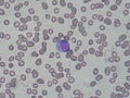
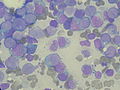 Bone marrow aspirate showing acute myeloid leukemia with Auer rods in several blasts
 -

-


References
edit- ^ Ackerman, G. Adolph (1950). "Microscopic and Histochemical Studies on the Auer Bodies in Leukemic Cells". Blood. 5 (9): 847–863. doi:10.1182/blood.V5.9.847.847. PMID 15434012.
- ^ Auer, John (1906). "Some hitherto undescribed structures found in the large lymphocytes of a case of acute leukaemia". American Journal of the Medical Sciences. 131 (6): 1002–1015. doi:10.1097/00000441-190606000-00008. ISSN 0002-9629. S2CID 71853154.
- ^ McCrae, Thomas (February 1905). "Acute lymphatic leukaemia with a report of five cases". British Medical Journal. 1 (2304): 404–408. doi:10.1136/bmj.1.2304.404. PMC 2319598. PMID 20761949.
- ^ Bain, Barbara (August 2011). "Auer rods or McCrae rods?". American Journal of Hematology. 86 (8): 689. doi:10.1002/ajh.21978. PMID 21761434.
External links
edit- Image at NIH/MedlinePlus
- Slides at wadsworth.org
- Image at University of Utah