In neuroanatomy, the maxillary nerve (V2) is one of the three branches or divisions of the trigeminal nerve, the fifth (CN V) cranial nerve. It comprises the principal functions of sensation from the maxilla, nasal cavity, sinuses, the palate and subsequently that of the mid-face,[1] and is intermediate, both in position and size, between the ophthalmic nerve and the mandibular nerve.[2]
| Maxillary nerve | |
|---|---|
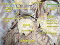
 Alveolar branches of superior maxillary nerve and pterygopalatine ganglion. | |
 Distribution of the maxillary and mandibular nerves, and the submaxillary ganglion. | |
| Details | |
| From | Trigeminal nerve |
| To | Infraorbital nerve, zygomatic nerve, palatine nerve, nasopalatine nerve, sphenopalatine ganglion |
| Identifiers | |
| Latin | nervus maxillaris |
| MeSH | D008442 |
| TA98 | A14.2.01.037 |
| TA2 | 6216 |
| FMA | 52724 |
| Anatomical terms of neuroanatomy | |
Structure edit
It begins at the middle of the trigeminal ganglion as a flattened plexiform band then it passes through the lateral wall of the cavernous sinus. It leaves the skull through the foramen rotundum, where it becomes more cylindrical in form, and firmer in texture. After leaving foramen rotundum it gives two branches to the pterygopalatine ganglion. It then crosses the pterygopalatine fossa, inclines lateralward on the back of the maxilla, and enters the orbit through the inferior orbital fissure. It then runs forward on the floor of the orbit, at first in the infraorbital groove and then in the infraorbital canal remaining outside the periosteum of the orbit. It then emerges on the face through the infraorbital foramen and terminates by dividing into inferior palpebral, lateral nasal and superior labial branches. The nerve is accompanied by the infraorbital branch of (the third part of) the maxillary artery and the accompanying vein.
Branches edit
Its branches may be divided into four groups, depending upon where they branch off: in the cranium, in the pterygopalatine fossa, in the infraorbital canal, or on the face.
In the cranium edit
- Middle meningeal nerve in the meninges
From the pterygopalatine fossa edit
- Zygomatic nerve (zygomaticotemporal nerve, zygomaticofacial nerve), through the Inferior orbital fissure
- Nasopalatine nerve, through the sphenopalatine foramen
- Posterior superior alveolar nerve
- Greater and lesser palatine nerves
- Pharyngeal nerve
In the infraorbital canal edit
On the face edit
- Inferior palpebral nerve
- Superior labial nerve
- Lateral nasal nerve
Function edit
The Maxillary nerve gives cutaneous branches to the face. It also carries parasympathetic preganglionic fibers (sphenopalatine) and postganglionic fibers (zygomatic, greater and lesser palatine and nasopalatine) to and from the pterygopalatine ganglion.
Additional images edit
-
 Maxillary nerve

See also edit
References edit
![]() This article incorporates text in the public domain from page 889 of the 20th edition of Gray's Anatomy (1918)
This article incorporates text in the public domain from page 889 of the 20th edition of Gray's Anatomy (1918)
Books edit
- Monkhouse, Stanley (2006). Cranial nerves - functional anatomy. Cambridge. ISBN 0-521-61537-2.
- Feneis, Heinz; Dauber, Wolfgang (2007). Pocket Atlas of Human Anatomy (5th ed.). Thieme. pp. 400–401.
External links edit
- MedEd at Loyola GrossAnatomy/h_n/cn/cn1/cnb2.htm
- cranialnerves at The Anatomy Lesson by Wesley Norman (Georgetown University) (VII)