
Size of this preview: 695 × 599 pixels. Other resolutions: 278 × 240 pixels | 557 × 480 pixels | 805 × 694 pixels.
Original file (805 × 694 pixels, file size: 114 KB, MIME type: image/jpeg)
| This is a file from the Wikimedia Commons. Information from its description page there is shown below. Commons is a freely licensed media file repository. You can help. |
Summary
| Description |
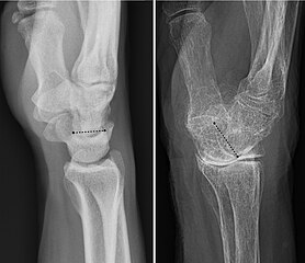
English: Projectional radiographs of a normal wrist (left image) and one (right image) with a dorsally tilted wrist joint due to wrist osteoarthritis, as well as osteoporosis. The the angle of the distal surface of the lunate bone is annotated. |
| Date | |
| Source |
|
| Author | Nevit Dilmen and Mikael Häggström |


Licensing
This file is licensed under the Creative Commons Attribution-Share Alike 3.0 Unported license.
- You are free:
- to share – to copy, distribute and transmit the work
- to remix – to adapt the work
- Under the following conditions:
- attribution – You must give appropriate credit, provide a link to the license, and indicate if changes were made. You may do so in any reasonable manner, but not in any way that suggests the licensor endorses you or your use.
- share alike – If you remix, transform, or build upon the material, you must distribute your contributions under the same or compatible license as the original.
File history
Click on a date/time to view the file as it appeared at that time.
| Date/Time | Thumbnail | Dimensions | User | Comment | |
|---|---|---|---|---|---|
| current | 14:12, 27 March 2017 | | 805 × 694 (114 KB) | Mikael Häggström | User created page with UploadWizard |
File usage
The following pages on the English Wikipedia use this file (pages on other projects are not listed):
Global file usage
The following other wikis use this file:
- Usage on vi.wikipedia.org