.png)
.png&action=edit&redlink=1)

Size of this preview: 683 × 600 pixels. Other resolutions: 273 × 240 pixels | 547 × 480 pixels | 875 × 768 pixels | 1,166 × 1,024 pixels | 2,332 × 2,048 pixels | 2,783 × 2,444 pixels.
Original file (2,783 × 2,444 pixels, file size: 2.95 MB, MIME type: image/png)
| This is a file from the Wikimedia Commons. Information from its description page there is shown below. Commons is a freely licensed media file repository. You can help. |
.png)
Summary
| Description |
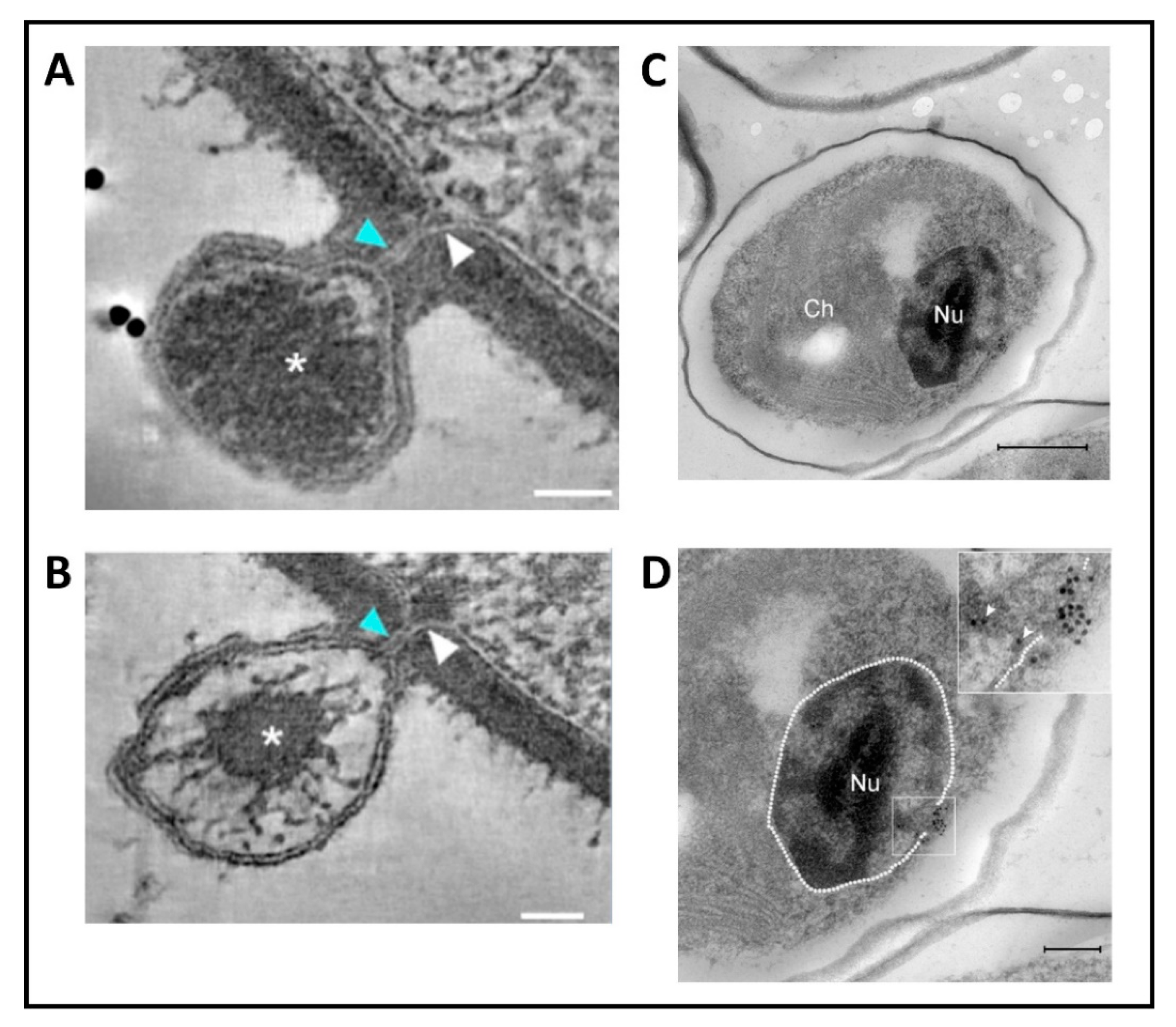
English: (A,B) PBCV-1 infected chlorella cells at 1.5–2 min p.i. were examined by Scanning-Transmission Electron Microscopy (STEM) tomography. The membrane-lined channel connecting the virus genome with the interior of the host is clearly visible. (A) A 7.8 nm tomographic slice from a 220 nm-thick STEM tomograph showing the close proximity between the viral and host internal membranes resulting from their convergence at the infection site. (B) A 5.2 nm tomographic slice from a different 220 nm STEM tomogram showing that part of the virus genome has been ejected into the cell. (C,D) Cells were infected with PBCV-1 for 6 min and then chemically fixed and thin sections were subjected to Electron Microscopy In Situ hybridization. (C) Low magnification of a cell illustrating dense viral DNA near the nucleus. (D) High magnification view of panel C. Note that viral DNA is probably entering the nucleus (white arrowheads in the inset). The nucleus contour is delineated with a white dashed line. The white and blue arrows (in A+B) point to the membranes that line the channel, the asterisk: viral DNA, Nu: nucleus, Ch; chloroplast, Scale bars: A, B: 50 nm; C: 500 nm; D: 200nm. |
| Date | |
| Source |
https://www.mdpi.com/viruses/viruses-12-00020/article_deploy/html/images/viruses-12-00020-g007.png at https://www.mdpi.com/1999-4915/12/1/20/htm (edit) Viruses 2020, 12(1), 20;doi:10.3390/v12010020 This article belongs to the Special Issue Viruses Ten-Year Anniversary. Licensee MDPI, Basel, Switzerland. This article is an open access article distributed under the terms and conditions of the Creative Commons Attribution (CC BY) license (https://creativecommons.org/licenses/by/4.0/). |
| Author | Provided by James L. Van Etten, Irina V. Agarkova, David D. Dunigan. Photohrapher(s): Milrot et al. |
| Other versions |
 |
Licensing
This file is licensed under the Creative Commons Attribution-Share Alike 4.0 International license.
- You are free:
- to share – to copy, distribute and transmit the work
- to remix – to adapt the work
- Under the following conditions:
- attribution – You must give appropriate credit, provide a link to the license, and indicate if changes were made. You may do so in any reasonable manner, but not in any way that suggests the licensor endorses you or your use.
- share alike – If you remix, transform, or build upon the material, you must distribute your contributions under the same or compatible license as the original.
File history
Click on a date/time to view the file as it appeared at that time.
| Date/Time | Thumbnail | Dimensions | User | Comment | |
|---|---|---|---|---|---|
| current | 19:26, 12 March 2021 | | 2,783 × 2,444 (2.95 MB) | Ernsts | Uploaded a work by Provided by James L. Van Etten, Irina V. Agarkova, David D. Dunigan. Photohrapher(s): Milrot ''et al.'' from https://www.mdpi.com/viruses/viruses-12-00020/article_deploy/html/images/viruses-12-00020-g007.png at https://www.mdpi.com/1999-4915/12/1/20/htm (edit) Viruses 2020, 12(1), 20;doi:10.3390/v12010020 This article belongs to the Special Issue Viruses Ten-Year Anniversary. Licensee MDPI, Basel, Switzerland. This article is an open access article distributed unde... |
File usage
The following pages on the English Wikipedia use this file (pages on other projects are not listed):
.png)