
Size of this preview: 800 × 398 pixels. Other resolutions: 320 × 159 pixels | 640 × 318 pixels | 1,024 × 509 pixels | 1,280 × 637 pixels | 3,519 × 1,750 pixels.
Original file (3,519 × 1,750 pixels, file size: 3.12 MB, MIME type: image/png)
| This is a file from the Wikimedia Commons. Information from its description page there is shown below. Commons is a freely licensed media file repository. You can help. |
Summary
| Description |
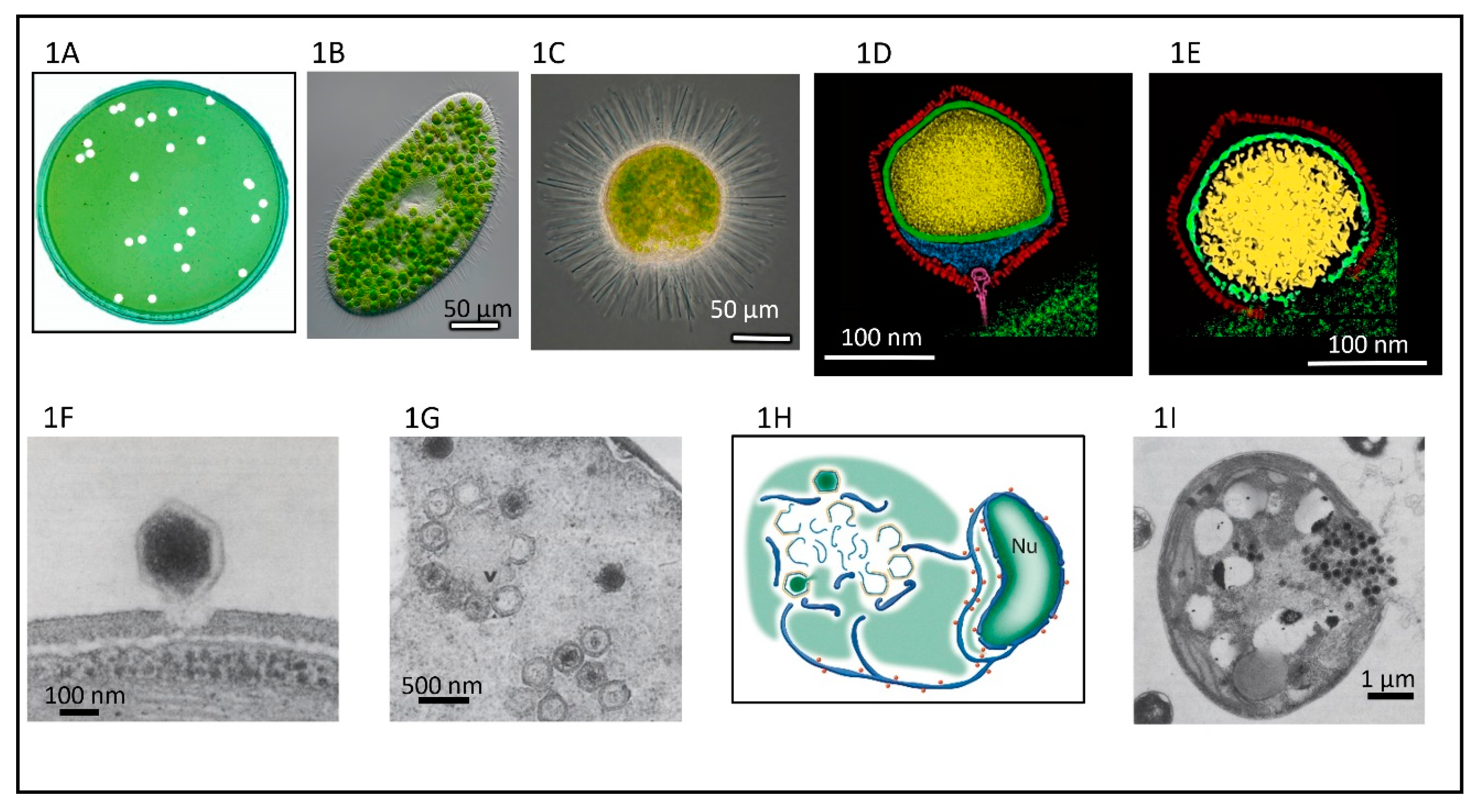
English: Chlorella cells and chlorovirus: Paramecium bursaria chlorella virus-1 (PBCV-1). (A) Plaques formed by PBCV-1 on a lawn of C. variabilis NC64A. (B) The ciliate Paramecium bursaria and its symbiotic chlorella cells. (C) The heliozoon Acanthocystis turfacea and its symbiotic chlorella cells. (D) Cross section of a five-fold averaged cryo-EM image of PBCV-1 reveals a long narrow cylindrical spike structure at one vertex and the viral internal membrane (green) surrounding the viral genome asymmetrically. (E) Cross-section of a five-fold averaged cryo-EM of PBCV-1 as the virus is getting ready to release its DNA into the host cell. (F) Attachment of PBCV-1 to the algal cell wall and degradation of the wall at the point of attachment. This occurs within 1–3 min post infection (p.i.). (G) PBCV-1 particles assemble in defined areas in the cytoplasm named virus assembly centers at ~5 h p.i. Note both DNA containing (dark centers) and empty capsids. (H) A model showing that the origin of the PBCV-1 internal membrane arises from nuclei-derived cisternae, which serve as precursors for the single bi-layered virus membrane. Note, the membrane serves as the template for the capsid structures to form virus particles. (I) Localized lysis of the cell plasma membrane and cell wall and release of progeny viruses at ~7 h p.i. Panels D and E are from the cover of J. Virology issue 17, 2012. |
| Date | |
| Source |
https://www.mdpi.com/viruses/viruses-12-00020/article_deploy/html/images/viruses-12-00020-g001.png at https://www.mdpi.com/1999-4915/12/1/20/htm (edit) Viruses 2020, 12(1), 20;doi:10.3390/v12010020 This article belongs to the Special Issue Viruses Ten-Year Anniversary. Licensee MDPI, Basel, Switzerland. This article is an open access article distributed under the terms and conditions of the Creative Commons Attribution (CC BY) license (https://creativecommons.org/licenses/by/4.0/). |
| Author | Provided by James L. Van Etten, Irina V. Agarkova, David D. Dunigan |
| Other versions |
.png) |
.png)
_PBCV-1.png)
_PBCV-1.png)
_PBCV-1@hostcell.png)
_PBCV-1_assembly.png)
_assembly_schema.png)
_PBCV-1_Lysis%2BRelease.png)
.png)
.png&action=edit&redlink=1)
.png)
Licensing
This file is licensed under the Creative Commons Attribution-Share Alike 4.0 International license.
- You are free:
- to share – to copy, distribute and transmit the work
- to remix – to adapt the work
- Under the following conditions:
- attribution – You must give appropriate credit, provide a link to the license, and indicate if changes were made. You may do so in any reasonable manner, but not in any way that suggests the licensor endorses you or your use.
- share alike – If you remix, transform, or build upon the material, you must distribute your contributions under the same or compatible license as the original.
File history
Click on a date/time to view the file as it appeared at that time.
| Date/Time | Thumbnail | Dimensions | User | Comment | |
|---|---|---|---|---|---|
| current | 19:26, 12 March 2021 | | 3,519 × 1,750 (3.12 MB) | Ernsts | Uploaded a work by Provided by James L. Van Etten, Irina V. Agarkova, David D. Dunigan from https://www.mdpi.com/viruses/viruses-12-00020/article_deploy/html/images/viruses-12-00020-g001.png at https://www.mdpi.com/1999-4915/12/1/20/htm (edit) Viruses 2020, 12(1), 20;doi:10.3390/v12010020 This article belongs to the Special Issue Viruses Ten-Year Anniversary. Licensee MDPI, Basel, Switzerland. This article is an open access article distributed under the terms and conditions of the Creativ... |
File usage
No pages on the English Wikipedia use this file (pages on other projects are not listed).
.png)