File:Simplified structure of the mammalian kidney lobe.svg

Original file(SVG file, nominally 364 × 333 pixels, file size: 63 KB)

Summary

Description
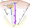
English: Numeric designations:
  1. Cortex
  2. Outer medulla
  3. Inner medulla
  4. Inner stripe
  5. Outer stripe
  6. Nephron
  7. Medullary ray
  8. Renal capsule
  9. Renal papilla

Color legends:

  • #9d18ec Collecting duct
  • #00f6e9 Connecting tubule
  • #8cabf8 Distal convoluted tubule
  • #134ef7 Distal straight tubule
  • #c3c3bc Intermediate tubule (loop of Henle)
  • #e7dc2e Proximal straight tubule
  • #dda330 Proximal convoluted tubule
Русский: Числовые обозначения:
  1. Корковое вещество
  2. Наружная зона мозгового вещества
  3. Внутренняя зона мозгового вещества
  4. Внутренняя полоса наружной зоны мозгового вещества
  5. Наружная полоса наружной зоны мозгового вещества
  6. Нефрон
  7. Мозговой луч
  8. Почечная капсула
  9. Почечный сосочек

Цветовые обозначения:

  • #9d18ec Collecting duct
  • #00f6e9 Connecting tubule
  • #8cabf8 Distal convoluted tubule
  • #134ef7 Distal straight tubule
  • #c3c3bc Intermediate tubule (loop of Henle)
  • #e7dc2e Proximal straight tubule
  • #dda330 Proximal convoluted tubule
Date
Source A. J. Davidson (15 January 2009). "Mouse kidney development". StemBook. doi:10.3824/STEMBOOK.1.34.1. PMID 20614633. URL: https://www.stembook.org/node/532
Author

Alan J. Davidson – author of original raster work

D6194c-1cc – vectorized work; modified glomerulus and macula densa position; lobe sectioning made rounded rather than linear; added numerical designations (instead of language-dependant texts); removed kidney image and color legends

Licensing

w:en:Creative Commons
attribution
This file is licensed under the Creative Commons Attribution 4.0 International license.
You are free:
  • to share – to copy, distribute and transmit the work
  • to remix – to adapt the work
Under the following conditions:
  • attribution – You must give appropriate credit, provide a link to the license, and indicate if changes were made. You may do so in any reasonable manner, but not in any way that suggests the licensor endorses you or your use.

Captions

Simplified structure of the mammalian kidney lobe

Items portrayed in this file

depicts

16 October 2023

image/svg+xml

File history

Click on a date/time to view the file as it appeared at that time.

Date/TimeThumbnailDimensionsUserComment
current06:26, 17 October 2023Thumbnail for version as of 06:26, 17 October 2023364 × 333 (63 KB)D6194c-1ccArrows made as paths (workaround to fix rendering bug)
22:52, 16 October 2023Thumbnail for version as of 22:52, 16 October 2023364 × 333 (61 KB)D6194c-1ccFixed arrows and glomerulus contours color
21:48, 16 October 2023Thumbnail for version as of 21:48, 16 October 2023364 × 333 (66 KB)D6194c-1ccUploaded a work by Alan J. Davidson – author of original raster work D6194c-1cc – vectorized work; modified glomerulus and macula densa position; lobe sectioning made rounded rather than linear; added numerical designations (instead of language-dependant texts); removed kidney image and color legends from A. J. Davidson (15 January 2009). "Mouse kidney development". StemBook. doi:10.3824/STEMBOOK.1.34.1. PMID 20614633. URL: https://www.stembook.org/node/532 with UploadWizard
The following pages on the English Wikipedia use this file (pages on other projects are not listed):

Global file usage

The following other wikis use this file:

Metadata