Size of this preview: 594 × 600 pixels. Other resolutions: 238 × 240 pixels | 475 × 480 pixels | 926 × 935 pixels.
Original file (926 × 935 pixels, file size: 1.56 MB, MIME type: image/jpeg)
| This is a file from the Wikimedia Commons. Information from its description page there is shown below. Commons is a freely licensed media file repository. You can help. |
Summary
| Description |
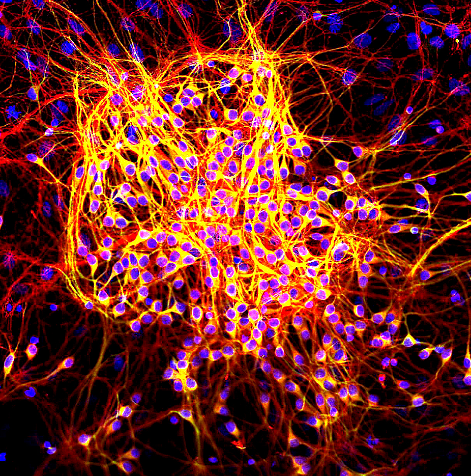
English: Neurons were grown in tissue culture and stained with antibody to MAP2 protein in green and MAP tau in red. MAP2 is found only in dendrites and perikarya, while tau is found not only in the dendrites and perikarya but also in axons. As a result axons appear red while the dendrites and perikarya appear yellow, due to superimposition of the red and green signals. DNA is shown in blue using the DAPI stain which highlights the nuclei. |
| Date | |
| Source | Own work |
| Author | GerryShaw |
Licensing
I, the copyright holder of this work, hereby publish it under the following license:
This file is licensed under the Creative Commons Attribution-Share Alike 4.0 International license.
- You are free:
- to share – to copy, distribute and transmit the work
- to remix – to adapt the work
- Under the following conditions:
- attribution – You must give appropriate credit, provide a link to the license, and indicate if changes were made. You may do so in any reasonable manner, but not in any way that suggests the licensor endorses you or your use.
- share alike – If you remix, transform, or build upon the material, you must distribute your contributions under the same or compatible license as the original.
File history
Click on a date/time to view the file as it appeared at that time.
| Date/Time | Thumbnail | Dimensions | User | Comment | |
|---|---|---|---|---|---|
| current | 20:01, 23 September 2014 | | 926 × 935 (1.56 MB) | GerryShaw | User created page with UploadWizard |
File usage
The following pages on the English Wikipedia use this file (pages on other projects are not listed):
Global file usage
The following other wikis use this file:
- Usage on bs.wikipedia.org
- Usage on ca.wikipedia.org
- Usage on da.wikipedia.org
- Usage on en.wiktionary.org
- Usage on ja.wikipedia.org
- Usage on ru.wikipedia.org
- Usage on sr.wikipedia.org