The allantois (/əˈlæntoʊɪs/ a-LAN-toe-iss;[1] pl.: allantoides or allantoises) is a hollow sac-like structure filled with clear fluid that forms part of a developing amniote's conceptus (which consists of all embryonic and extraembryonic tissues). It helps the embryo exchange gases and handle liquid waste.
| Allantois | |
|---|---|
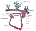
 Diagram illustrating a chicken egg in its 9th day with all extraembryonic membranes | |
 Allantois and other fetal membranes shown at end of second month of human pregnancy | |
| Details | |
| Pronunciation | /əˈlæntɔɪs/, /əˈlæntoʊɪs/ |
| Days | 16 |
| Identifiers | |
| Latin | allantois |
| MeSH | D000482 |
| TE | E6.0.1.2.0.0.2 |
| Anatomical terminology | |
The allantois, along with the amnion, chorion, and yolk sac (other extraembryonic membranes), identify humans and other mammals, birds, and reptiles as amniotes. These extraembryonic membranes that form the embryo have aided amniotes in the transition from aquatic to terrestrial environments.[2] Fish and amphibians are anamniotes, and lack the allantois. In mammals the extraembryonic membranes are known as the fetal membranes.
Function edit
This sac-like structure, whose name is Greek for sausage (from ἀλλαντοειδής allantoeidḗs, in reference to its shape when first formed)[3] is primarily involved in nutrition and excretion, and is webbed with blood vessels. The function of the allantois is to collect liquid waste from the embryo, as well as to exchange gases used by the embryo.
In mammals edit
In mammals excluding egg-laying monotremes, the allantois is one of the fetal membranes, and is part of and forms an axis for the development of the umbilical cord.
While the function of the allantois remains conserved, there is a divergence in the characteristics of the allantois among mammalian species. Structural variations of the allantois include differences in size and shape.[4]
In mammals, the emergence of the allantois varies during early development. In humans, the allantois appears during the presomitic stages of mid-gastrulation. The mouse allantois appears before the first somite pair as well; however, it occurs shortly after gastrulation. In both pigs and rabbits, the allantois arises at early somite stages.[4]
The human allantois is a caudal out-pouching of the yolk sac, which becomes surrounded by the mesodermal connecting stalk known as the body-stalk. The vasculature of the body-stalk develops into umbilical arteries that carry deoxygenated blood to the placenta.[5] It is externally continuous with the proctodeum and internally continuous with the cloaca. The embryonic allantois becomes the fetal urachus, which connects the fetal bladder (developed from cloaca) to the yolk sac. The urachus removes nitrogenous waste from the fetal bladder.[6] After birth the urachus becomes obliterated as the median umbilical ligament.
The mouse allantois consists of mesodermal tissue, which undergoes vasculogenesis to form the mature umbilical artery and vein.[7]
In marsupials edit
In most marsupials, the allantois is avascular, having no blood vessels, but still serves the purpose of storing nitrogenous (NH3) waste. Also, most marsupial allantoises do not fuse with the chorion. An exception is the allantois of the bandicoot, which has a vasculature, and fuses with the chorion. Koalas and wombats also demonstrate allantoic fusion with the chorion.[8]
In reptiles and birds edit

The structure first evolved in reptiles and birds as a reservoir for nitrogenous waste, and also as a means for oxygenation of the embryo. Oxygen is absorbed by the allantois through the egg shell.
In most reptiles and birds, the allantois consists of extraembryonic endoderm enclosed in mesodermal tissue. A chorioallantoic membrane (CAM) is later formed from the chorion and allantois.[2]
Clinical significance edit
During the third week of embryonic development, the allantois protrudes into the area of the urogenital sinus. During fetal development, the allantois becomes the urachus, a duct between the bladder and the yolk sac. A patent allantois can result in a urachal cyst.
Because the allantois can be cultured ex vivo, it has utility as a model system for studying the formation of blood vessels as well as considerable usefulness in drug screening.[9]
Additional images edit
-
 Section through the embryo.
 -
 Diagram showing later stage of allantoic development with commencing constriction of the yolk-sac.
 -
 Opened uterus with cat fetus in midgestation: 1 umbilicus, 2 amniotic sac (chorion and amnion), 3 allantois.


References edit
- ^ Jones, Daniel (October 6, 2011). Cambridge English Pronouncing Dictionary. Cambridge University Press. ISBN 9780521765756 – via Google Books.
- ^ a b Chuva de Sousa Lopes, Susana M.; Roelen, Bernard A. J.; Lawson, Kirstie A.; Zwijsen, An (2022-12-05). "The development of the amnion in mice and other amniotes". Philosophical Transactions of the Royal Society B: Biological Sciences. 377 (1865): 20210258. doi:10.1098/rstb.2021.0258. ISSN 0962-8436. PMC 9574641. PMID 36252226.
- ^ "Allantois". dictionary.com.
- ^ a b Hassan, Waad; Viebahn, Christoph (May 2017). "A correlative study of the allantois in pig and rabbit highlighting the diversity of extraembryonic tissues in four mammalian species, including mouse and man: HASSAN AND VIEBAHN". Journal of Morphology. 278 (5): 600–620. doi:10.1002/jmor.20657.
- ^ Moore, Keith; Persaud, T.V.N.; Torchia, Mark G. (2016). The Developing Human: Clinically Oriented Embryology (10th ed.). Philadelphia: Elsevier. p. 58. ISBN 978-0-323-31338-4.
- ^ First AID for the USMLE Step 1 2008
- ^ Downs, KM (1998). "The Murine Allantois". Curr Top Dev Biol. 39: 1–33. doi:10.1016/s0070-2153(08)60451-2. PMID 9475996.
- ^ Carter, Anthony M. (July 2021). "The role of mammalian foetal membranes in early embryogenesis: Lessons from marsupials". Journal of Morphology. 282 (7): 940–952. doi:10.1002/jmor.21140. ISSN 0362-2525.
- ^ Arora, Ripla; Papaioannou, Virginia E. (2012-09-27). "The murine allantois: a model system for the study of blood vessel formation". Blood. 120 (13): 2562–2572. doi:10.1182/blood-2012-03-390070. ISSN 0006-4971. PMC 3460680. PMID 22855605.